Connective Tissue Graft: The Wall That Protects Your Teeth
Leggi in Italiano
In brief — A connective tissue graft restores keratinized gum tissue, rebuilding the biological barrier that protects teeth from gum recession. It’s the surgical gold standard, with results that stay stable for decades.
Every day, millions of people lose gum tissue millimeter by millimeter without noticing.
Periodontal disease — which includes gingivitis and periodontitis — is probably the single most common infectious disease group there is. And often the worst cases are silent, with few or no symptoms at all.
By the time someone notices there’s a problem, the root is usually already exposed, sensitive, vulnerable.
But there’s a way to rebuild that natural protection and stop the disease from progressing.
Recognize the problem
Gum tissue isn’t just a cosmetic lining. It’s a wall. A biological barrier that keeps bacteria away from the tooth root and absorbs the forces of brushing and chewing. When that wall thins or pulls back, the tooth loses its first line of defense. And it’s a strong mechanical defense.
The science is clear: tooth surfaces with less than 2 millimeters of protective gum tissue show inflammation in 80% of cases, even with impeccable hygiene (Lang & Löe, 1972). It’s not about brushing better. It’s about structure.
Why gums recede
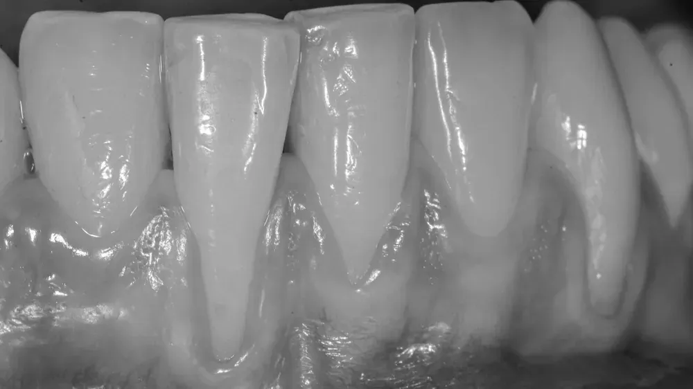
The causes are many: overly aggressive brushing, a genetically thin biotype, orthodontic treatment, restorations placed too deep, bruxism. Whatever the trigger, the outcome is always the same: the root gets exposed, becomes sensitive to hot and cold, and the risk of root decay rises because the root has no enamel to protect it.
The problem is that gum recession doesn’t stop on its own. Left untreated, it tends to progress. And every tenth of a millimeter lost is a tenth of a millimeter less protection.
The solution: rebuilding the wall (and removing the risk factors)
Connective tissue grafting is the technique science recognizes as the gold standard for restoring lost gum tissue (Chambrone et al., Cochrane 2018). The principle is simple: harvest a small fragment of tissue from an area of the patient’s own mouth where that same tissue is abundant — and regenerates naturally, with no consequences — and place it where it’s needed, beneath the existing gum.
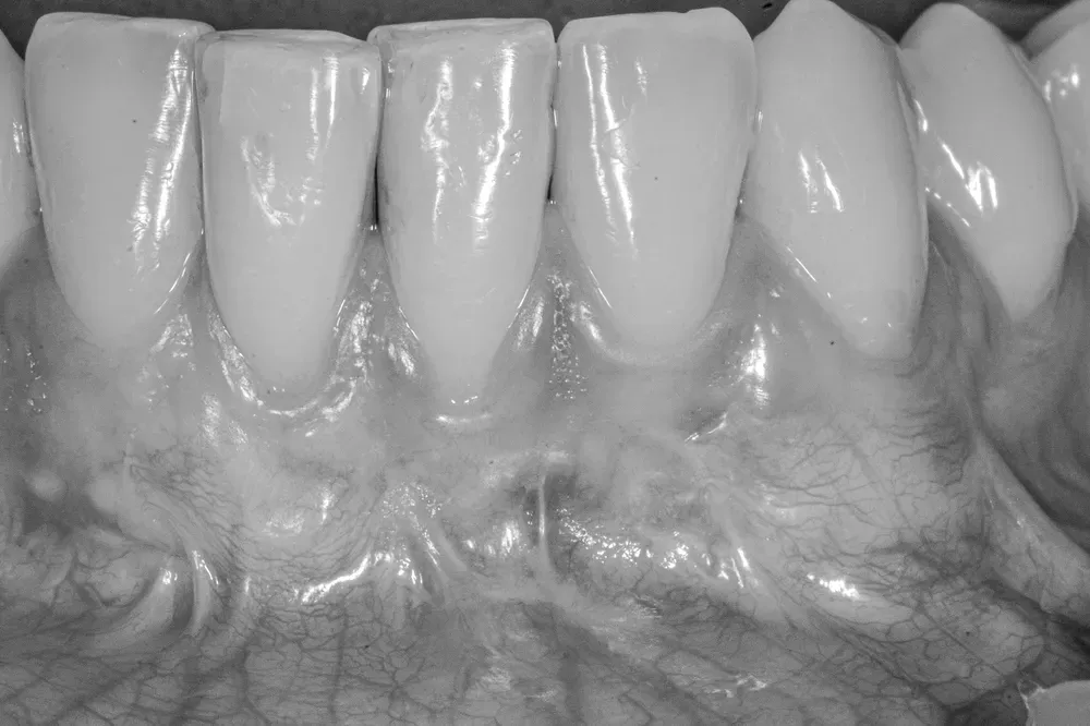
This graft doesn’t just cover the exposed root. It does far more: it induces the formation of new keratinized gum tissue, the tough, dense tissue that forms the real protective barrier. In other words, it rebuilds the wall.
In recent years, xenogeneic collagen membranes have reached the market, designed to avoid a second surgical site on the palate. They’re convenient, they reduce postoperative pain, but they’re not equivalent. A meta-analysis of 16 randomized trials and 632 patients, published by Koppolu and colleagues in 2026, measured average root coverage 11–13 percentage points lower with membranes than with autologous grafts, at 6 and 12 months (Koppolu et al., 2026). The gap is even wider for keratinized tissue gain. The palate is still an awkward thing to explain to a patient — but it’s still the option that works best.
The scientific background
The scientific literature documents results that few other procedures in dentistry can claim. Coverage of the exposed root reaches 85–99% of cases (Oates et al., AAP Consensus 2015). But the more important number is about durability.
A 10-year study found that sites treated with connective tissue grafting were 39 times more likely to maintain complete coverage than sites treated without a graft (Cairo et al., 2023). And longer follow-ups, out to 20 years, confirm the result stays stable over time (Petsos et al., 2020).
But the coverage percentage, on its own, doesn’t tell the whole story. In 2025, a systematic review of 41 preclinical and 43 clinical studies went looking for what actually happens under the microscope: autologous connective tissue grafting remains the only technique that documents a true new connective attachment, with fibers oriented perpendicular to the root — not a simple epithelial re-adhesion destined to fail at the first trauma (Shanbhag et al., 2025).
Which, when you think about it, makes sense.
Once it has taken, it can only grow.
We’re not talking about a temporary fix. We’re talking about a reconstruction that lasts decades.
How the wall works
The difference between keratinized gum tissue (thick and firm) and oral mucosa (delicate, thin, fragile) lies in the microscopic structure. Keratinized gum tissue has a protective layer similar to skin, with tightly connected epithelial cells and a dense, resistant connective tissue firmly attached to the underlying teeth and bone (Histology, Oral Mucosa - NCBI).
The mucosa lining areas without attached gum tissue is, by contrast, thin, mobile, permeable. It doesn’t effectively withstand daily stress (like brushing), nor does it help keep bacteria from infiltrating toward the root when the keratinized band is thin.
Connective tissue grafting turns this fragile mucosa into attached, keratinized gum tissue. The transplanted tissue carries with it the genetic information to produce a thick epithelial layer and a deep layer firmly attached to the underlying tissues, and within a few weeks it induces the formation of a new protective barrier, biologically identical to the original.
When a graft is actually needed
Not every recession requires surgery. The decision depends on several factors: how deep the recession is, how thick the remaining tissue is, whether there’s sensitivity or an esthetic concern, and above all the risk of progression.
But once the indication is there, waiting doesn’t pay off. The further a recession progresses, the more complex the recovery becomes. And some situations — like fillings or crowns that extend below the gumline — make surgery almost mandatory to prevent chronic inflammation (Stetler & Bissada, 1987).
The result you can expect
A properly performed procedure restores the tooth’s natural protection. Sensitivity disappears, esthetics improve, and above all, that silent process of progressive loss — which would otherwise have continued unchecked — comes to a stop.
And what does the patient actually see in the mirror? Two systematic reviews, from 2025 and 2026, published in Periodontology 2000, compared the clinician’s judgment against the judgment of the person looking in the mirror every morning. For single recessions, professional esthetic scores and patient-reported satisfaction converge in most cases (Cairo et al., 2025). For multiple recessions — 32 randomized trials, over 1,000 patients analyzed — average root coverage tops out at 82.6%, yet satisfaction still climbs to 8.59 out of 10: one more confirmation that the clinical result and the perceived one don’t always match, and that the second one often matters more (Stefanini et al., 2026).
The grafted tissue integrates completely with the surrounding tissue. After healing, no one can tell the rebuilt gum from the original. And check-ups years later confirm the wall holds.
The next step
If you notice your gums are receding, if your teeth look longer than they used to, if you feel sensitivity to cold in areas that never bothered you before, don’t wait for it to get worse.
A thorough evaluation can determine whether your case needs treatment or just careful monitoring. Either way, knowing beats not knowing.
Ask your dentist.
References
- Koppolu P, et al. Comparative Effectiveness of Autogenous Connective Tissue Grafts and Xenogeneic Soft Tissue Substitutes for Multiple Gingival Recessions: A Systematic Review and Meta-Analysis. Medicina (Kaunas). 2026;62(2):366. doi:10.3390/medicina62020366. PMID: 41752765.
- Shanbhag S, Stødle IH, Lie SA, Sanz M, Verket A. Histological Outcomes of Root Coverage Procedures: A Systematic Review With Meta-Analysis. J Periodontal Res. 2026;61:9-29. doi:10.1111/jre.70043. PMID: 41017258.
- Cairo F, et al. Clinician- and Patient-Reported Outcomes Following the Surgical Treatment of Single Gingival Recession Defects: A Systematic Review. Periodontol 2000. 2025. doi:10.1111/prd.12641. PMID: 40693671.
- Stefanini M, Mounssif I, Figuero E, Zucchelli G, Sculean A, Cosgarea R. Esthetical and Patient-Reported Outcomes After Root Coverage Procedures for Multiple Gingival Recessions: A Systematic Review and Meta-Analysis. Periodontol 2000. 2026. doi:10.1111/prd.70050. PMID: 42130372.
FAQ
Is a gum graft procedure painful?
How long does full healing take?
Are the results of a gum graft permanent?
Where is the tissue for the graft harvested from?
Are there alternatives to grafting tissue from the palate?
Can I eat normally after the procedure?
Can a gum graft fail?
Can multiple teeth be treated in a single session?
Cite this article
Ready for theses, slides and papers. If you cite it on a web page, make the URL a link so readers can reach the source.
Bruschi E. Connective Tissue Graft: The Wall That Protects Your Teeth [Internet]. Frosinone (Italy): Bonebenders; 2025 [updated 2026 Jul 13; cited 2026 Oct 25]. Available from: https://bonebenders.com/en/blog/connective-tissue-graft-gum-recession-treatment/ Bruschi, E. (2025, December 5). Connective Tissue Graft: The Wall That Protects Your Teeth. Bonebenders. https://bonebenders.com/en/blog/connective-tissue-graft-gum-recession-treatment/
@misc{bruschi2025connective,
  author       = {Bruschi, Ernesto},
  title        = {Connective Tissue Graft: The Wall That Protects Your Teeth},
  year         = {2025},
  month        = dec,
  howpublished = {Bonebenders},
  url          = {https://bonebenders.com/en/blog/connective-tissue-graft-gum-recession-treatment/},
  note         = {Accessed 2026-10-25}
} Go deeper
Espansione crestale (split crest) →Allargare la cresta senza trapianto — tecnica ERE e bonebending
Need a professional opinion?
Book an appointment at Dr. Bruschi's practice in Frosinone. First visit includes full diagnosis and personalised treatment plan.
Stay Updated
New articles on periodontology, implantology and oral surgery — delivered to your inbox.
Comments
Loading comments...
Leave a comment