480 men, 20 years, no toothbrushes: Löe's Sri Lanka study
Leggi in Italiano
In brief — Between 1970 and 1990 Harald Löe followed 480 workers on two tea plantations in Sri Lanka: no toothbrush, no toothpaste, no periodontist available, no periodontal therapy. Plaque, calculus and gingivitis were everywhere. And what happened to these men? Around 8% had lost every tooth by 45. In 81% the periodontitis advanced slowly; in 11% it never went beyond gingivitis, with no loss of the supporting tissues at all. Individual susceptibility entered periodontology here and stayed. Grades A, B and C in the 2018 classification are the direct descendants of those three groups.
Eleven men in a hundred never touched a toothbrush for twenty years. And even so, by the age of forty-five they had lost a little over one millimetre of periodontal attachment. In the same population, on the same diet, doing the same work, carrying the same amount of plaque on their teeth, eight in a hundred had no teeth left at forty-five.
Why did some of them develop the disease and others not? Why do identical conditions fail to produce identical effects in every person?
Why Sri Lanka?
Harald Löe was looking for a population nobody had ever treated — truly nobody. There was a precise methodological reason. Every longitudinal study run in Europe or the United States was measuring a disease already altered by treatment, by hygiene instruction, by extractions carried out for other reasons. Nobody had ever seen the natural history of periodontitis, because in the West somebody always intervened.
He found it on two tea plantations in the Sri Lankan highlands. In 1970 Löe, Ånerud, Boysen and Smith examined 480 male labourers aged between 14 and 31, and published the study design and the baseline data in the Journal of Periodontal Research (Löe et al., 1978). None of those men had ever used a toothbrush. None had ever seen a hygienist. None had ever received adequate periodontal therapy — nor even an extraction.
One further detail makes the cohort ideal. That population was practically free of caries. Every missing tooth had been lost to periodontal disease. Counting teeth therefore became a direct measure of the outcome of this disease, rather than of decay.
In parallel, the same research group was following a set of Norwegian students and academics in Oslo who received regular dental care — the “control” group. Two populations at opposite ends of the same continuum, measured with the same protocol for twenty years.
The first answer was the wrong one
In 1978, in the Journal of Periodontology, the researchers published the comparison between the two cohorts before the age of forty. The Norwegians reached forty with a little over 1.5 mm of mean attachment loss and a rate of 0.08 mm per year on interproximal surfaces. The Sri Lankans, at the same age, stood at 4.50 mm, losing 0.30 mm per year (Löe et al., 1978).
The conclusion of that paper was that, left alone, the periodontal lesion progresses at a fairly constant and continuous rate.
Eight years later the same authors wrote the opposite. On the same study population.
Three kinds of periodontitis
The 1986 paper in the Journal of Clinical Periodontology is the one everybody cites, and rightly so. On the basis of interproximal attachment loss and tooth loss, Löe and colleagues split the cohort into three subpopulations (Löe et al., 1986):
- Rapid progression (RP), around 8%. At 35: 9 mm of mean attachment loss and 12 teeth already gone. At 40: 20 teeth missing. At 45: nothing left. The annual rate ranged between 0.1 and 1.0 mm.
- Moderate progression (MP), around 81%. Four millimetres at 35, seven at 45. Tooth loss began after thirty. This is the overwhelming majority of the population, and its curve is the one that most resembles what we see in the surgery.
- No progression (NP), around 11%. Under one millimetre at 35, with an annual rate between 0.05 and 0.09 mm — inside the noise of the measurement itself. Plaque and calculus in abundance, gingivitis practically everywhere, and nothing that went any further.
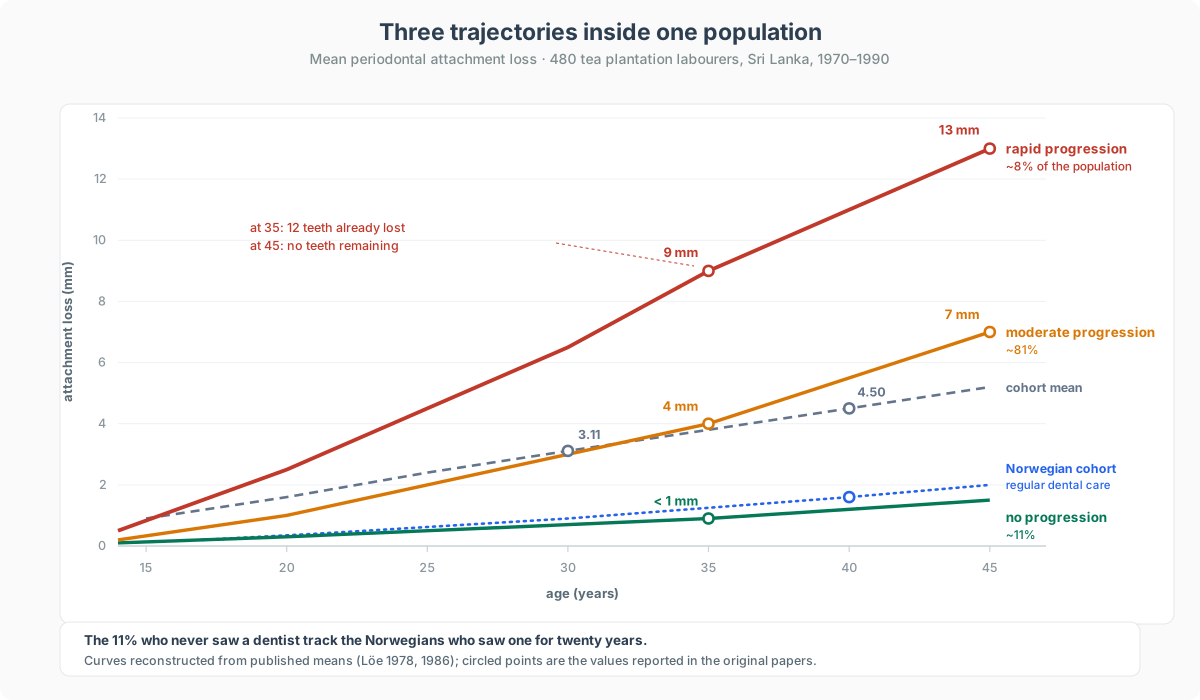
Reading the chart, the mean of the tea workers’ cohort sits far above the Norwegian control group. And the control group essentially coincides with the “no progression” curve of the experimental cohort. Remarkable. Conclusive.
The Sri Lankan 11%, with no care of any kind and having never seen a dentist, ends up where the Norwegians ended up after twenty years of regular dental attention.
That 11% changed the discipline
Until then periodontitis was explained by a linear equation: more plaque, more disease. It was the model that had replaced the older idea of pyorrhoea as constitutional degeneration, and it looked settled because it worked on the aggregate numbers of many studies of the period.
The 11% broke it. Those men carried enormous deposits of plaque and calculus on every surface, gingival inflammation on virtually every gingival surface, and thirty years of uninterrupted exposure to all of it. Their periodontal attachment stayed exactly where it was.
The unavoidable conclusion is that the host response decides the outcome. The same bacterial insult produces nine millimetres of loss in one man and none in another, even within the same ethnicity, on the same diet, in the same trade. The cohort’s microbiota, incidentally, held nothing exotic: Prevotella intermedia in 76% of subjects, Porphyromonas gingivalis in 40%, Aggregatibacter actinomycetemcomitans in 15%, distributed across healthy and diseased sites much as in industrialised countries (Preus et al., 1995).
In 1992 the group also published its data on gingival recession in the two cohorts, and the figures are striking: by forty, 100% of the Sri Lankans had recession, against far lower rates among the Norwegians, where it was confined to buccal surfaces (Löe et al., 1992).
A few considerations
You will read almost everywhere that the study demonstrated the role of plaque, calculus, gingivitis and smoking as the engines of progression. The twenty-year risk factor analysis published by Neely, Holford, Löe, Ånerud and Boysen in 2001 says something else. In the final adjusted model, age, gingival index, calculus index and follow-up time were significant, all at p < 0.0001. The plaque index, smoking history and betel nut consumption were not significantly associated with attachment loss over time (Neely et al., 2001).
In the rest of the literature smoking is a solid risk factor. Here its effect was probably masked by the other risk factors, present on a massive scale. In the 1991 paper on calculus, labourers who both smoked and chewed betel had significantly higher calculus scores than those who did only one of the two, and teeth carrying calculus lost attachment significantly faster than teeth that stayed clear (Ånerud et al., 1991).
On the final outcome — losing the tooth — betel comes back. In the 2005 analysis of 455 subjects, tooth loss depended on the interaction between attachment loss and betel nut use, which amplified its effect (Neely et al., 2005). A factor can be irrelevant on an intermediate measure and decisive on the clinical outcome that counts.
What is left of this study?
In 2018 the joint AAP/EFP World Workshop rewrote the classification of periodontal diseases, adding grade alongside stage: A for slow progression, B for moderate, C for rapid, with risk factors used as grade modifiers (Tonetti, Greenwell & Kornman, 2018).
Periodontology has understood that the individual condition of the patient is essential to understanding the disease and its treatment.
The new classification dropped the old split between chronic and aggressive periodontitis, which described two diseases where there is one moving at different speeds. And it is why, in the surgery, the prognostic estimate weighs more than the snapshot of the moment.
The limits
The 480 men of 1970 were 161 original participants at the 1985 examination, and 154 in the cohort used for the twenty-year risk factor analysis. The attrition in those numbers is enormous.
On a tea plantation, people often leave the follow-up feet first. It would be interesting to know the causes of those deaths as well.
All male. One occupational group, one diet, one socioeconomic setting. No genetic measurement: in 1970 “susceptibility” was inferred from the trajectory, never measured directly. Saying that the 11% had a favourable immune response is an inference, not a laboratory finding.
And the three-group structure was invisible during the first eight years of observation. It emerged only once the cohort had been followed long enough — which ought to make all of us cautious when we extrapolate a rate of progression, on our own single and debatable judgement, from two radiographs a year apart.
The legacy
Löe went to those plantations to describe the outcome of untreated disease. He came back with something different and more useful: the proof that the same disease, under the same conditions, produces very different outcomes in each person.
From then on the clinical question became how does this patient respond to the plaque and calculus he has. Every time we stand in front of a severe case and ask ourselves why him, we are thinking the way the great Löe thought.
References
- Löe H, Ånerud A, Boysen H, Smith M. The natural history of periodontal disease in man. Study design and baseline data. J Periodontal Res. 1978;13(6):550-62. doi:10.1111/j.1600-0765.1978.tb00209.x · PMID: 153395
- Löe H, Ånerud A, Boysen H, Smith M. The natural history of periodontal disease in man. Tooth mortality rates before 40 years of age. J Periodontal Res. 1978;13(6):563-72. doi:10.1111/j.1600-0765.1978.tb00210.x · PMID: 153396
- Löe H, Ånerud A, Boysen H, Smith M. The natural history of periodontal disease in man. The rate of periodontal destruction before 40 years of age. J Periodontol. 1978;49(12):607-20. doi:10.1902/jop.1978.49.12.607 · PMID: 282430
- Löe H, Ånerud A, Boysen H, Morrison E. Natural history of periodontal disease in man. Rapid, moderate and no loss of attachment in Sri Lankan laborers 14 to 46 years of age. J Clin Periodontol. 1986;13(5):431-45. doi:10.1111/j.1600-051x.1986.tb01487.x · PMID: 3487557
- Ånerud A, Löe H, Boysen H. The natural history and clinical course of calculus formation in man. J Clin Periodontol. 1991;18(3):160-70. doi:10.1111/j.1600-051x.1991.tb01128.x · PMID: 2061415
- Löe H, Ånerud A, Boysen H. The natural history of periodontal disease in man: prevalence, severity, and extent of gingival recession. J Periodontol. 1992;63(6):489-95. doi:10.1902/jop.1992.63.6.489 · PMID: 1625148
- Preus HR, Ånerud A, Boysen H, Dunford RG, Zambon JJ, Löe H. The natural history of periodontal disease. The correlation of selected microbiological parameters with disease severity in Sri Lankan tea workers. J Clin Periodontol. 1995;22(9):674-8. doi:10.1111/j.1600-051x.1995.tb00825.x · PMID: 7593696
- Neely AL, Holford TR, Löe H, Ånerud A, Boysen H. The natural history of periodontal disease in man. Risk factors for progression of attachment loss in individuals receiving no oral health care. J Periodontol. 2001;72(8):1006-15. doi:10.1902/jop.2001.72.8.1006 · PMID: 11525431
- Neely AL, Holford TR, Löe H, Ånerud A, Boysen H. The natural history of periodontal disease in humans: risk factors for tooth loss in caries-free subjects receiving no oral health care. J Clin Periodontol. 2005;32(9):984-93. doi:10.1111/j.1600-051X.2005.00797.x · PMID: 16104963
- Tonetti MS, Greenwell H, Kornman KS. Staging and grading of periodontitis: framework and proposal of a new classification and case definition. J Periodontol. 2018;89 Suppl 1:S159-S172. doi:10.1002/JPER.18-0006 · PMID: 29926952
FAQ
Who ran the study on the natural history of periodontal disease?
How many took part, and for how long were they followed?
What are the RP, MP and NP groups?
Does the study show that plaque does not matter?
Did smoking emerge as a risk factor in that cohort?
Cite this article
Ready for theses, slides and papers. If you cite it on a web page, make the URL a link so readers can reach the source.
Bruschi E. 480 men, 20 years, no toothbrushes: Löe's Sri Lanka study [Internet]. Frosinone (Italy): Bonebenders; 2026 [cited 2026 Oct 25]. Available from: https://bonebenders.com/en/blog/loe-sri-lanka-natural-history-periodontitis/ Bruschi, E. (2026, September 4). 480 men, 20 years, no toothbrushes: Löe's Sri Lanka study. Bonebenders. https://bonebenders.com/en/blog/loe-sri-lanka-natural-history-periodontitis/
@misc{bruschi2026lanka,
  author       = {Bruschi, Ernesto},
  title        = {480 men, 20 years, no toothbrushes: Löe's Sri Lanka study},
  year         = {2026},
  month        = sep,
  howpublished = {Bonebenders},
  url          = {https://bonebenders.com/en/blog/loe-sri-lanka-natural-history-periodontitis/},
  note         = {Accessed 2026-10-25}
} Go deeper
Espansione crestale (split crest) →Allargare la cresta senza trapianto — tecnica ERE e bonebending
Need a professional opinion?
Book an appointment at Dr. Bruschi's practice in Frosinone. First visit includes full diagnosis and personalised treatment plan.
Stay Updated
New articles on periodontology, implantology and oral surgery — delivered to your inbox.
Comments
Loading comments...
Leave a comment