Wisdom Teeth: When to Remove Them and When Not To
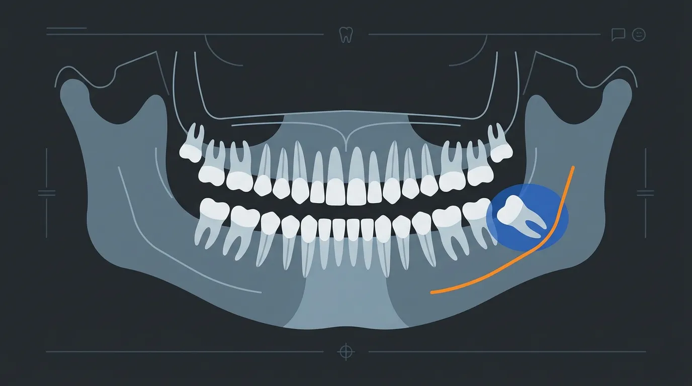
One of the few procedures where the right question is not “how it’s done” but “whether it should be done at all.”
Wisdom teeth are probably the topic on which patients arrive at the office with the most preconceived opinions. The friend who “was in agony for a week.” The TikTok video where all four are removed under sedation. The previous dentist who said “let’s keep an eye on them” for fifteen years without ever actually looking.
The reality is both simpler and more complicated. Simpler, because the indications for extraction are few and clear. More complicated, because the decision not to extract is equally clinical — and often harder to explain.
What we are talking about
Third molars — the “eights” — are the last teeth to erupt, usually between the ages of 17 and 25. In a modern mandible, there is often insufficient space to accommodate them. The result is a tooth that remains partially or totally trapped in the bone: impacted. This occurs in up to 73% of young European adults. Not all of them cause problems. But when they do, they do so decisively.
The possible positions are many — mesioangular, distoangular, horizontal, vertical — and the Winter classification, together with the Pell-Gregory classification, tells us a great deal about surgical difficulty and complication risk. This is not an academic detail: the position of the tooth is the first prognostic factor for the procedure.
When to remove: the clear indications
The indications for extraction are codified. They are not opinions.
Recurrent pericoronitis — inflammation of the soft tissue that partially covers the tooth. Non-restorable decay, whether of the wisdom tooth or the adjacent second molar. Root resorption of the second molar. Follicular cysts or other associated pathology. Documented orthodontic reasons. Abscesses that cannot be resolved with conservative treatment.
When any of these conditions is present, extraction is not a choice: it is the therapy. Delaying it does not improve the prognosis — it worsens it, because surgical complications increase with age and with disease progression.
When to leave it alone: the prophylactic dilemma
And here the most controversial chapter opens. The asymptomatic, fully impacted wisdom tooth with no radiographic pathology: should it be removed or left alone?
The NICE guidelines (2000, reaffirmed in 2020) are explicit: prophylactic extraction of asymptomatic, disease-free third molars is not recommended. The rationale is that surgery exposes a patient who currently has no problem to surgical risks.
Recent literature, however, adds nuance. Hounsome et al. (2020), in a systematic review commissioned by the British NIHR, built a long-term economic model and found that the rate of extraction for teeth initially left in place ranges from 5.5% to 31.4% at five years. Their model suggests that prophylactic extraction in patients in their twenties may be the most cost-effective strategy — with an incremental cost of just £55.71 and a gain in quality of life, albeit a small one.
This is not an invitation to remove everything from everyone. It is an invitation not to settle the question with a dogma in either direction. The thoughtful clinician evaluates each individual case: position, angulation, relationship with the inferior alveolar nerve, age, general health status, and history of previous inflammation.
The real risks — and how to reduce them
Every surgical procedure carries risks. Honesty lies in quantifying them, not in hiding them and not in exaggerating them either.
The complication that concerns most is injury to the inferior alveolar nerve — the nerve that provides sensation to the lower lip and chin. The reported incidence in the literature is 0.5–5%, with most cases resolving spontaneously within 6 months. Lingual nerve injury is rarer (0.1–2%) but potentially more bothersome for the patient. Dry socket — the early loss of the blood clot — affects 1–4% of cases and is managed with local dressings.
Bailey et al. (2020), in the Cochrane review on surgical techniques for mandibular third molars — 62 trials, 4,643 patients — found that the evidence on which technique is superior remains of low quality. The only indication with any degree of support is the use of PRF/PRP in the post-extraction socket to reduce the incidence of dry socket. Beyond that: the surgeon’s skill matters more than the technique chosen.
CBCT: is it necessary or not?
The panoramic radiograph remains the first-line imaging study. But when it shows signs of proximity between the wisdom tooth roots and the mandibular canal — white line interruption, canal deviation, root darkening — the question of CBCT arises.
The answer is counterintuitive. Clé-Ovejero et al. (2017), in a meta-analysis published in JADA, compared the incidence of nerve injury in patients imaged with three-dimensional CT versus panoramic radiograph alone: no statistically significant difference (RR 0.96; 95% CI: 0.50–1.85; p = 0.91). CBCT does not reduce the risk of injury. But it is absolutely useful — because it changes the surgical plan. Knowing whether the root is buccal or lingual relative to the nerve canal, whether an intact cortical plate exists, or whether the nerve runs through the roots modifies the surgical strategy. And in certain cases, it indicates coronectomy.
Coronectomy: the alternative that works
Coronectomy is perhaps the most underrated procedure in oral surgery. It consists of removing only the crown of the impacted tooth, leaving the roots in situ. It seems counterintuitive — leaving roots inside the bone — but the numbers speak clearly.
Two recent meta-analyses (Peixoto et al., 2023: 42 studies, 3,095 patients; Kang et al., 2025: 34 studies, 7,115 teeth) converge on the same result: coronectomy reduces the risk of inferior alveolar nerve injury by 86–90% compared to complete extraction (OR 0.14 and RR 0.1, both with p < 0.001). It also reduces dry socket. The trade-off is a failure rate of 2.79% and a reoperation probability of 3.63% (Kang et al.).
In practice: out of 100 high-risk patients, coronectomy prevents nerve injury in approximately 4–5 who would have experienced it with complete extraction, at the cost of 3 reoperations. The balance is favourable, and the AAOMS guidelines recommend it when CBCT documents an intimate relationship between the roots and the nerve.
What to expect after the procedure
Swelling is the norm, not the exception. It peaks at 48–72 hours and resolves within 5–7 days. Pain follows a predictable pattern: the peak arrives 5–6 hours after the procedure, when the anaesthesia wears off. The combination of ibuprofen 400 mg + paracetamol 500–1000 mg is the most effective therapy (NNT 2.3) — better than opioids, with fewer side effects. Ice in the first 24 hours helps.
Soft diet for 3–5 days. No aspirin. No vigorous rinsing in the first 24 hours — the clot must be protected, not dislodged. Sutures are either resorbable or removed after 7–10 days. Full soft tissue healing takes 2–3 weeks.
Feeling anxious? That’s normal — and manageable.
The difference between a surgeon and Google
Many websites sell generic reassurance: “It’s a routine procedure.” But every wisdom tooth is different. A mesioangular third molar in class I-A is a 10-minute operation. A horizontal one in class III-C with roots embracing the nerve is a procedure that requires planning, advanced imaging, and the ability to decide — in the operating room — whether to complete the extraction or convert to coronectomy.
In Frosinone, at our practice, every impacted third molar extraction begins with a recent panoramic radiograph. If radiographic signs warrant it, we proceed with CBCT. The clinical decision — extract, perform coronectomy, or monitor — is made based on data, not on one-size-fits-all protocols.
Because the right question is not “will you remove my wisdom tooth?” The right question is “in my specific case, is it worth it?” And the honest answer, sometimes, is no.
References
- Bailey E, Kashbour W, Shah N, Worthington HV, Renton TF, Coulthard P. Surgical techniques for the removal of mandibular wisdom teeth. Cochrane Database Syst Rev. 2020;7(7):CD004345.
- Kang FW, Yuan XR, Li GC, Yang YFZ, Zhang XM, Hou GY. Coronectomy in Lower Third Molar Surgery: A Systematic Review and Meta-Analysis. J Oral Maxillofac Surg. 2025;83(5):601-615.
- Peixoto AO, Bachesk AB, Leal MOCD, Jodas CRP, Machado RA, Teixeira RG. Benefits of Coronectomy in Lower Third Molar Surgery: A Systematic Review and Meta-analysis. J Oral Maxillofac Surg. 2023;82(1):73-92.
- Hounsome J, Pilkington G, Mahon J, Boland A, Beale S, Kotas E, et al. Prophylactic removal of impacted mandibular third molars: a systematic review and economic evaluation. Health Technol Assess. 2020;24(30):1-116.
- Clé-Ovejero A, Sánchez-Torres A, Camps-Font O, Gay-Escoda C, Figueiredo R, Valmaseda-Castellón E. Does 3-dimensional imaging of the third molar reduce the risk of experiencing inferior alveolar nerve injury owing to extraction? A meta-analysis. J Am Dent Assoc. 2017;148(8):575-583.
FAQ
When should a wisdom tooth be removed?
What are the risks of wisdom tooth extraction?
What is coronectomy and when is it used?
Is a CT scan (CBCT) needed before wisdom tooth extraction?
How long is recovery after wisdom tooth extraction?
Go deeper
Espansione crestale (split crest) →Allargare la cresta senza trapianto — tecnica ERE e bonebending
Need a professional opinion?
Book an appointment at Dr. Bruschi's practice in Frosinone. First visit includes full diagnosis and personalised treatment plan.
Stay Updated
New articles on periodontology, implantology and oral surgery — delivered to your inbox.
Comments
Loading comments...
Leave a comment