Oral Surgery in Frosinone: What We Treat and How We Do It
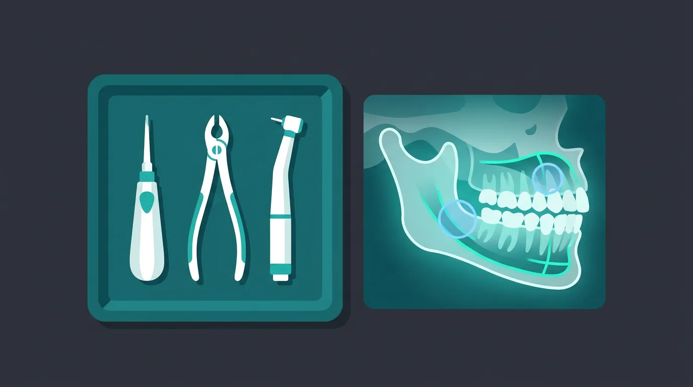
Every procedure begins before you touch the patient. It begins with a diagnosis.
Oral surgery is the branch of dentistry that deals with everything requiring a surgical act in the oral cavity and its associated structures. Put that way, it sounds straightforward. It is not — because the mouth is a surgical field of a few square centimeters where nerves, arteries, maxillary sinuses, and structures that do not forgive imprecision all converge.
At our practice in Frosinone, oral surgery is not an ancillary service. It is one of the areas where we invest the most — in training, technology, and protocols. This article explains what we treat, how we do it, and why certain choices that look like details are actually the decisions that make the difference between a procedure that goes well and one that goes perfectly.
Complex extractions and wisdom teeth
Extracting a tooth is not always a straightforward gesture. Curved roots, ankylosis, proximity to vital structures, teeth fractured below the gum line: each of these conditions turns a “simple” extraction into a procedure that demands planning.
Wisdom teeth deserve a separate discussion — and a dedicated article — because the most important decision is not how to remove them but whether to remove them. The indications are clear: recurrent pericoronitis, non-restorable caries, resorption of the adjacent molar, cysts, orthodontic reasons. But the asymptomatic impacted third molar is another chapter, and the debate between prophylactic extraction and monitoring is far from settled.
When the roots of a lower wisdom tooth are intimately related to the inferior alveolar nerve, coronectomy — removal of the crown alone while preserving the roots — reduces the risk of nerve injury by 86–90%. This is not a surgical surrender: it is an evidence-based strategy confirmed by two meta-analyses in the Journal of Oral and Maxillofacial Surgery (2023 and 2025).
Jaw cysts
Odontogenic cysts — dentigerous, radicular, residual, keratocysts — are pathological cavities lined by epithelium that grow slowly within the bone. Often asymptomatic for years, they are discovered incidentally on a panoramic radiograph. But “asymptomatic” does not mean “harmless”: an untreated cyst erodes bone, displaces roots, and in rare cases can undergo transformation.
Treatment depends on the nature and size of the lesion. Enucleation — complete removal of the cyst along with its epithelial lining — is the gold standard for lesions of manageable size. Marsupialization — opening the cyst and allowing prolonged drainage — is the strategy for large cysts, where immediate enucleation would risk damaging adjacent structures or creating an excessively large bony defect.
In both cases: everything that is removed goes to pathology. Always. A biopsy is not optional — it is the only way to obtain a definitive diagnosis.
Apicoectomy
When an endodontically treated tooth continues to cause problems — a periapical granuloma that does not resolve, an impossible endodontic retreatment, a persistent apical lesion — apicoectomy is the surgical route.
The root apex is accessed through the bone, the terminal portion of the root is resected (the apical 3 mm, where most of the endodontic anatomical complexity is concentrated), a retrograde cavity is prepared, and it is sealed with a biocompatible cement — MTA or Biodentine. The long-term success rate ranges from 85 to 95%, depending on technique and diagnosis.
Preoperative CBCT, here, is not optional. It identifies the number and configuration of root canals, the relationship with adjacent structures, and the extent of the lesion — information that two-dimensional radiography simply cannot provide.
Biopsies and soft tissue lesions
A white patch on the mucosa. An ulcer that has not healed in three weeks. A submucosal nodule. In most cases, it is benign. But “in most cases” is not “always” — and the only way to know for certain is a biopsy.
The dentist is often the first to see what the patient does not notice. Leukoplakia, lichen planus, an early-stage carcinoma. An incisional or excisional biopsy is a simple, quick surgical procedure performed under local anesthesia — and it can save a life.
Frenulectomy
The frenulum — upper labial or lingual — can be short, fibrous, or inserted too close to the gingival margin or the alveolar ridge. The consequences range from a midline diastema to restricted tongue mobility (ankyloglossia) to gingival recession.
A frenulectomy is a minor procedure — 15–20 minutes under local anesthesia — but timing matters. In a child with severe ankyloglossia, early intervention improves breastfeeding and speech development. In adults, it is often combined with periodontal surgery or orthodontics.
Tori and exostoses
Tori — mandibular and palatal — and vestibular exostoses are benign bony outgrowths. They are not pathological, but they can become a problem: they interfere with removable prostheses, make oral hygiene difficult, ulcerate under prosthetic loading, or simply worry the patient.
Surgical recontouring is an elective procedure: the excess bone is removed, the ridge is reshaped, and the site is sutured. Healing takes 10–14 days. Nothing heroic — but it requires a steady hand and a calibrated osteotomy to avoid damaging the underlying cortical bone.
Oroantral communication management
Extraction of an upper premolar or molar can occasionally create an oroantral communication — a direct connection between the oral cavity and the maxillary sinus. If not recognized and managed immediately, it becomes a chronic fistula.
Primary closure with a pedicled flap — buccal (Rehrmann) or palatal — is the most reliable technique. Success depends on immediate diagnosis: if the surgeon tests for the communication at the end of the extraction (Valsalva maneuver, gentle probing), it can be closed in the same session. If ignored, the problem becomes both surgical and sinusal.
Pain management: pre-emptive, not reactive
Post-surgical pain management is where much of the literature separates modern surgery from what was done twenty years ago. The concept is simple: pain is prevented, not chased.
The pain peak arrives 5–6 hours after surgery, when the local anesthesia wears off. If you wait until it hurts to take pain medication, you are already behind. The pre-emptive protocol calls for:
- Expected mild pain: ibuprofen 400 mg before the anesthesia wears off
- Expected moderate pain: ibuprofen 400 mg + paracetamol 1000 mg in combination
- Expected severe pain: bupivacaine at end of surgery (analgesia 8–12 hours) + NSAID upon awakening
The ibuprofen + paracetamol combination has an NNT of 2.3 — the best among all oral analgesics. Better than opioids, and without their side effects.
Afraid of surgery? That is normal — and manageable.
Imaging: see before you cut
Every surgical procedure starts with an image. A panoramic radiograph is the first step: an overview of the arches, sinuses, and mandibular canal. But it is not always enough.
CBCT comes into play when the third dimension is needed: the root-to-nerve relationship in impacted wisdom teeth, the three-dimensional extent of a cyst, planning apicoectomies on multi-rooted teeth, evaluation of oroantral communications. It is not a routine exam — it is an exam prescribed when two-dimensional imaging leaves questions unanswered.
At our practice in Frosinone, CBCT is on site. There is no need to send the patient elsewhere, no weeks of waiting. Imaging and surgery in the same facility, often during the same diagnostic appointment.
Why the oral surgeon is not interchangeable
Oral surgery is a medical act. It is not a mechanical gesture. Behind every extraction lies a risk assessment, a choice of technique, a plan B if plan A does not work.
Choosing an oral surgeon means choosing someone who knows how to read a CBCT, who understands the difference between a dentigerous cyst and a keratocyst, who knows when an extraction should be converted to a coronectomy, who manages pain with evidence-based protocols rather than prescribing opioids “just to be safe.”
At our practice in Frosinone, every surgical decision is made on data. Not on habit.
References
- Bailey E, Kashbour W, Shah N, Worthington HV, Renton TF, Coulthard P. Surgical techniques for the removal of mandibular wisdom teeth. Cochrane Database Syst Rev. 2020;7(7):CD004345.
- Kang FW, Yuan XR, Li GC, Yang YFZ, Zhang XM, Hou GY. Coronectomy in Lower Third Molar Surgery: A Systematic Review and Meta-Analysis. J Oral Maxillofac Surg. 2025;83(5):601-615.
- Peixoto AO, Bachesk AB, Leal MOCD, Jodas CRP, Machado RA, Teixeira RG. Benefits of Coronectomy in Lower Third Molar Surgery: A Systematic Review and Meta-analysis. J Oral Maxillofac Surg. 2023;82(1):73-92.
- Malamed SF. Management of pain and anxiety. In: Hupp JR, Ellis E, Tucker MR, editors. Contemporary Oral and Maxillofacial Surgery. 7th ed. St. Louis: Elsevier; 2019. p. 39-60.
FAQ
What oral surgery procedures are performed at the Frosinone office?
How is pain managed after oral surgery?
Is a CT scan required before oral surgery?
How long does it take to heal after oral surgery?
What are the risks of oral surgery?
Go deeper
Espansione crestale (split crest) →Allargare la cresta senza trapianto — tecnica ERE e bonebending
Need a professional opinion?
Book an appointment at Dr. Bruschi's practice in Frosinone. First visit includes full diagnosis and personalised treatment plan.
Stay Updated
New articles on periodontology, implantology and oral surgery — delivered to your inbox.
Comments
Loading comments...
Leave a comment