Horizontal Bone Defects: The Meta-Analysis on 1,400 Implants
In brief — A meta-analysis of 1,400 implants confirms the efficacy of the ridge split: 98.1% survival and 3.3 mm of bone gain. The technique is first-line for moderate horizontal defects, with a manageable risk profile and lower costs than bone grafting.
Sintesi (IT) — La meta-analisi su 1.400 impianti conferma l’efficacia della ridge split: 98,1% di sopravvivenza e 3,3 mm di guadagno osseo. La tecnica è la prima scelta per i deficit orizzontali moderati, con un profilo di rischio gestibile e costi contenuti rispetto al bone grafting.
…revealing which technique delivers predictable results
The technique every practice should master
What can I say, except: “good — the literature seems to be heading exactly where we’ve always liked it to go…”
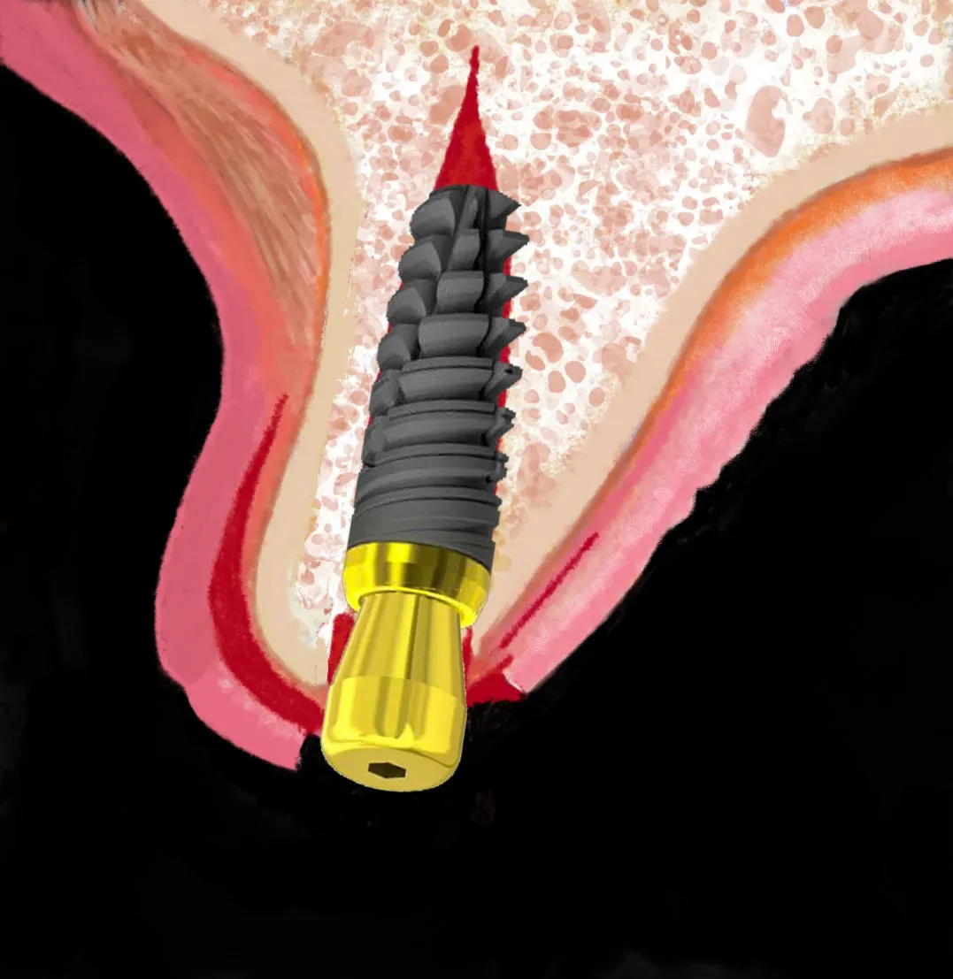
Horizontal bone defects are one of the most frequent challenges in modern implantology. When a patient presents with an alveolar ridge that is too narrow, the clinician faces a crucial choice: which augmentation technique delivers the most predictable results with the lowest risk of complications?
An answer comes from the largest meta-analysis ever conducted on the Alveolar Ridge Split (ARS) technique, published by Lin et al. in BMC Oral Health in 2023. The study analysed 25 clinical investigations (including one of the many studies by Bruschi [my father] and co-workers) carried out over 13 years, evaluating more than 1,400 implants to provide definitive evidence on the efficacy of the procedure.
The research that brings clarity
Lin et al.’s meta-analysis is the first large-scale systematic study dedicated exclusively to what the authors call Alveolar Ridge Split (ARS). The researchers applied strict inclusion criteria, selecting only studies with at least 10 patients that reported quantitative measurements of horizontal bone gain and implant survival data.
The methodology followed the PRISMA guidelines and the search was registered in the PROSPERO database, ensuring transparency and reproducibility. The authors analysed partially or fully edentulous patients undergoing implant surgery with ARS, including different technical variants and anatomical sites.
The numbers that shift the clinical perspective
For the first time, the meta-analysis provides reliable pooled data on ridge split efficacy. The analysis of 14 studies showed a mean horizontal bone gain of 3.348 mm (95% CI: 2.533–4.163 mm), with an observed range between 1.6 and 5.3 mm. This gain is clinically significant for implant placement in most cases of moderate deficit.
Even more relevant is the implant survival rate that emerged from the analysis of 17 studies: 98.1% (95% CI: 96.9%–98.9%). This figure, with a range from 93.9% to 100%, is perfectly comparable to the survival rates of standard implantology, confirming the long-term predictability of the technique.
The consistency of results across different studies, conducted in different centres and with multiple technical variants, reinforces the validity of the evidence. Bone gain proved reproducible regardless of the specific protocol used, suggesting an intrinsic robustness of the procedure.
Not an isolated finding: the literature converges
A single number proves nothing. A direction does.
And here the direction is clear. A few months before Lin, Al Haydar, Kang and Momen-Heravi published in the International Journal of Oral & Maxillofacial Implants (2023) an even larger meta-analysis: 35 studies, 4,446 implants, 98.17% survival and a mean horizontal gain of 3.06 mm. Numbers that overlap with Lin’s, but on a sample three times the size. The detail that really interests me, though, is another one: there was no statistical difference between sites treated with a graft and those left to heal without any biomaterial (3.06 mm versus 3.06 mm — essentially identical). Translation: the bone chips bought at a premium price, in a ridge split, almost never move the needle. This is exactly the philosophy that separates bonebending from traditional bone regeneration I have been repeating for years.
Vorovenci and colleagues (Biomedical Reports, 2024) then did the most honest thing one can do: place the techniques side by side. Osseodensification 2.15 mm, GBR 4.04 mm, ridge split 3.66 mm. Guided regeneration gains a few tenths more — but at what price in time, cost and morbidity? The ridge split sits a hair behind, with no donor site and no membranes. For moderate defects, the arithmetic of the risk–benefit ratio leaves little room for doubt.
Documented competitive advantages
The comparison with alternative augmentation techniques reveals distinctive advantages of the ridge split. The surgical time is significantly reduced compared with bone block grafting, eliminating the need for a donor site and consequently lowering patient morbidity. The possibility of placing the implant(s) immediately in many cases also shortens overall treatment time, unlike what happens with standardised all-on-X solutions.
Contained costs are a further competitive advantage, especially important in a market increasingly attentive to cost-effectiveness. The elimination of expensive biomaterials and the reduction of operating times translate into economic benefits for both clinician and patient.
From a biological standpoint, the technique exploits the intrinsic regenerative potential of bone, preserving local vascularisation and promoting a more physiological healing than procedures requiring heterologous or autologous grafts. This approach rests on the principles of natural bone remodelling.
This is not conference theory. Azadi and co-workers (Oral and Maxillofacial Surgery, 2025), in a meta-analysis dedicated solely to bone expansion without any graft, reported a 100% implant survival rate when the implant was placed simultaneously with the expansion. Bone, left to do its job, rarely disappoints. It is also confirmation that immediate placement, in the right bone defect, is not a gamble but often the more sensible choice.
Risk management and case selection
The analysis of complications documented across the studies reveals a manageable risk profile that nonetheless demands careful case selection. Buccal plate fractures are the most frequent complication, particularly when residual bone thickness is below 3 mm.
Author’s note: true, but this depends on inappropriate instruments and technique (which is why I always recommend keeping osteosynthesis screws or pins in the practice — it almost never happens, but it is better to be ready…).
According to this study’s data, dehiscences and exposures occur in 2.3–20.13% of cases, while minor infections affect 4.7–11.54% of patients. Transient paraesthesias, reported in 9.09–19.23% of cases, generally resolve spontaneously without permanent sequelae.
Residual bone thickness emerges as the most important predictive factor for success of the procedure. With thicknesses below 3 mm, the risk of fractures increases significantly, making a two-stage approach or alternative techniques preferable. Bone density is another critical factor: type D1 bone, particularly dense, presents greater technical difficulties than D2–D3 types.
Author’s note: it is no news that bone thickness is decisive. It is decisive in osteo-mucosal expansion techniques too. Yet, with the right technique, one can dare…
Evidence-based indications and contraindications
The data allow precise indications for the ridge split to be defined. Ideal candidates present horizontal bone defects of 3–4 mm with a residual thickness of at least 3 mm. Type D2–D3 bone, more frequent in the maxilla, offers optimal conditions for the procedure. Post-operative patient compliance is fundamental to prevent complications.
Relative contraindications include severe atrophies with less than 3 mm of residual bone, the very dense cortical bone typical of some mandibular sites, and systemic conditions that compromise bone healing. Posterior mandibular sites require particular attention, often benefiting from a two-stage approach to reduce fracture risk.
Author’s note: true, but this depends on inappropriate instruments and technique. With the right tools, even these sites and this type of bone can be regenerated with osteo-mucosal expansion techniques.
Anatomical site significantly influences results. The maxilla, with its more spongy and vascularised bone, offers more favourable conditions than the mandible. The greater elasticity of maxillary bone allows wider expansions with a lower fracture risk.
Author’s note: this too is broadly true but, once again, it depends on inappropriate instruments and technique. Otherwise it would be hard to explain why, for example, my personal database holds more cases of mandibular than maxillary expansion.
Limitations and future perspectives
Despite the encouraging results, the meta-analysis has limitations that condition its interpretation. The heterogeneity of surgical techniques, post-operative protocols and patient-selection criteria limits the generalisability of the findings. The predominance of observational studies lowers the level of evidence compared with randomised controlled trials.
The variability in follow-up, from 6 months to 10 years, prevents uniform assessments of long-term stability. Many studies lack data beyond 5 years — a critical window for evaluating bone and implant stability over time.
The bone gain capped at 3.3 mm, though clinically significant, is lower than the 4.25 mm achievable on average with bone block grafting. This limitation restricts the ridge split to cases of moderate deficit, excluding the severe atrophies that require more substantial augmentation.
Implications for clinical practice
The evidence reshapes the clinical approach to horizontal bone defects. The ridge split positions itself as first choice in moderate defects, offering a favourable risk–benefit ratio when applied with appropriate selective criteria. Accurate case selection is the key factor in optimising results.
Implementing standardised protocols for pre-operative assessment, surgical technique and post-operative follow-up is essential to replicate the literature’s results. The use of piezoelectric techniques can reduce surgical trauma and improve predictability, particularly in borderline cases.
There is also a hot-off-the-press finding: Gokila Vani, Bansal and colleagues (Journal of Oral Implantology, 2025) compared, in a controlled clinical trial on atrophic sites, piezoelectric surgery and screw expanders against conventional drilling. Implant stability (ISQ), insertion torque and the inflammatory marker TNF-α proved comparable across techniques. The reading is simple: expansion, when guided by modern instruments and a codified technique like bonebending, penalises neither the bone nor the implant. What flattens is the learning curve, not the biology.
Author’s note: as a daily user, diamond surgical discs can be superior.
Specific training and the technical learning curve must not be underestimated. The procedure requires surgical experience to manage anatomical variables and intra-operative complications, making adequate training necessary before clinical implementation. Modern instruments, however, help flatten the curve.
Evidence-based conclusions
Lin et al.’s meta-analysis provides robust evidence on the efficacy of the osteo-mucosal expansion technique — even when performed with varied and inconsistent methods — as a valid alternative for horizontal bone augmentation.
The mean gain of 3.3 mm and the 98.1% survival rate make it a predictable procedure when applied with appropriate selective criteria.
Author’s note: as a daily user and educator of osteo-mucosal expansion techniques, I should also stress that the possible complications reported by some of the selected studies are surely attributable to the specific surgical technique used. With the techniques and instruments we propose, the chance of complications is not only far lower than with the “Bonedriller” approaches but genuinely rare. What truly matters is knowing how to tell apart research that confirms already-known principles and real clinical innovation.
Frequently asked questions
🔍 How do I assess whether a case is suitable for Ridge Split?
The main criteria are: residual bone thickness ≥ 3 mm, a horizontal deficit of 3–4 mm, type D2–D3 bone, and the absence of systemic conditions that compromise healing. CBCT is fundamental to evaluate thickness, density and local anatomy. Avoid the technique with thicknesses < 3 mm or very dense bone (D1) without specific experience.
⚙️ What is the difference between Ridge Split and osteo-mucosal expansion?
The traditional Ridge Split uses chisels and osteotomes to separate the cortical plates. The Bonebenders osteo-mucosal expansion uses specific instruments (diamond discs) that allow greater control, less trauma, and the possibility of working even on dense bone and borderline thicknesses. The end result is similar, but the technique is more predictable and less traumatic.
📊 Is 98.1% survival realistic in everyday practice?
Yes, if the selection criteria are respected. The meta-analysis figure reflects well-conducted studies with appropriate case selection. In daily practice, success depends on the clinician’s experience, patient selection, post-operative compliance and adequate follow-up. Do not apply the technique to every bone defect — selection is fundamental.
🚑 How do I manage a buccal cortical plate fracture?
Do not panic. Options: 1) Osteosynthesis with screws/pins if the fragment is viable and of adequate size; 2) Removal of the fragment and guided bone regeneration; 3) Two-stage approach — healing then implant in a second phase. Always keep osteosynthesis materials in the practice. Most fractures are managed successfully.
⏱️ Immediate placement or better a two-stage approach?
Immediate when: residual thickness ≥ 4 mm, D2–D3 bone, controlled expansion without fractures, primary stability > 35 Ncm. Two-stage when: thickness 3–4 mm, very dense D1 bone, patient risk factors, lack of adequate primary stability. The two-stage approach is always safer in borderline cases.
💰 How much can this type of procedure cost the patient?
The economic advantage is significant: no expensive biomaterials, no donor site, reduced surgical times. The cost is typically 30–50% lower than GBR with a bone block. For the patient: less time in the chair, fewer appointments, faster healing. The saving allows quality treatment to be offered to more patients.
🔄 What do I do if the expansion is not sufficient?
Options: 1) Combine with GBR — expansion plus membranes/biomaterials for residual defects; 2) A second expansion procedure after 6–8 weeks; 3) Convert to an alternative technique (bone block). Never force the expansion beyond biological limits. A combined approach beats a complication.
🧬 Should I use biomaterials, or is spontaneous healing enough?
The Bonebenders philosophy favours spontaneous healing whenever possible. In a well-executed Ridge Split, the clot is sufficient if kept stable. Biomaterials are useful when: the gap is > 2 mm, volumetric maintenance is needed, or the patient is at risk. Nature has an extraordinary regenerative power — we just have to create the optimal conditions.
References
- Lin Y, Li G, Xu T, Zhou X, Luo F. The efficacy of alveolar ridge split on implants: a systematic review and meta-analysis. BMC Oral Health. 2023;23(1):894. doi:10.1186/s12903-023-03643-2 · PMID: 37986181
- Al Haydar B, Kang P, Momen-Heravi F. Efficacy of horizontal alveolar ridge expansion through the alveolar ridge split procedure: a systematic review and meta-analysis. Int J Oral Maxillofac Implants. 2023;38(6):1083-1096. doi:10.11607/jomi.9972 · PMID: 38085739
- Vorovenci A, Drafta S, Petre A. Horizontal ridge augmentation through ridge expansion via osseodensification, guided bone regeneration and ridge-split: systematic review and meta-analysis of clinical trials. Biomed Rep. 2024;21(4):139. doi:10.3892/br.2024.1827 · PMID: 39161939
- Azadi A, Hazrati P, Tizno A, Rezaei F, Akbarzadeh Baghban A, Tabrizi R. Bone expansion as a horizontal alveolar ridge augmentation technique: a systematic review and meta-analysis. Oral Maxillofac Surg. 2025;29(1):32. doi:10.1007/s10006-025-01335-5 · PMID: 39808204
- Gokila Vani SU, Bansal M, Srikrishna S, Shilpi S, Taslim A, Rashika M, et al. Horizontal augmentation via ridge splitting and expansion for implant placement in atrophic sites: a prospective non-randomized controlled trial. J Oral Implantol. 2025;51(6):513-521. doi:10.1563/aaid-joi-D-24-00240 · PMID: 41365336
Are you a colleague interested in these concepts? If you like, join our community at the bottom of the page.
References
Looking for a specialist?
Rigenerazione Ossea a Frosinone →Il Metodo Bonebenders: espansione osteo-mucosa senza innesti
Need a professional opinion?
Book an appointment at Dr. Bruschi's practice in Frosinone. First visit includes full diagnosis and personalised treatment plan.
Stai valutando un impianto dentale?
Ho scritto una guida in 8 capitoli che spiega tutto quello che un paziente dovrebbe sapere prima di sedersi in poltrona. Niente marketing — solo fatti, casi studio e una checklist per fare le domande giuste.
Scarica la guidaStay Updated
New articles on periodontology, implantology and oral surgery — delivered to your inbox.
Comments
Loading comments...
Leave a comment