In brief — Porphyromonas gingivalis does not stay in the periodontal pocket. It invades host cells, spreads cell-to-cell through cytoskeleton bridges, and persists in a dormant state. It exploits CD47 and thrombospondin-1 to dodge immune clearance. Its gingipains degrade key immune proteins and can breach the blood-brain barrier. That intracellular persistence is what connects it to Alzheimer’s disease, gastric and colorectal cancer, rheumatoid arthritis — and, now, fatty liver.
Sintesi (IT) — Il Porphyromonas gingivalis non resta nelle tasche parodontali. Invade le cellule ospiti, si trasmette da cellula a cellula attraverso ponti del citoscheletro e persiste in stato dormiente. Sfrutta CD47 e trombospondina-1 per evadere il sistema immunitario. Le sue gingipaine degradano proteine immunitarie chiave e possono alterare la barriera ematoencefalica. La persistenza intracellulare spiega le connessioni con Alzheimer, cancro gastrico e colorettale, artrite reumatoide e steatosi epatica.
Porphyromonas gingivalis lives in the depths of the periodontal pocket. It might look like a static situation. It isn’t.
When periodontal disease advances, the aggressive species take over. P. gingivalis is one of them. More than that: it’s the single species capable of destabilising the whole pocket ecosystem — the keystone pathogen. And what it does looks less like a plain infection and more like a military invasion.
The microenvironment of the gingival pocket
The bottom of the pocket is dark, warm, poorly oxygenated. Pathogenic bacteria find shelter even in the tartar deposits they helped to build — a fortress raised with their own means.
Without proper therapy, these species multiply exponentially. They are Gram-negative anaerobes: they draw energy from fermentation and anaerobic respiration, metabolising proteins and amino acids in the absence of oxygen. The by-products — fatty acids, hydrogen, methane, amines — are part of the chemical arsenal that assaults the surrounding tissue.
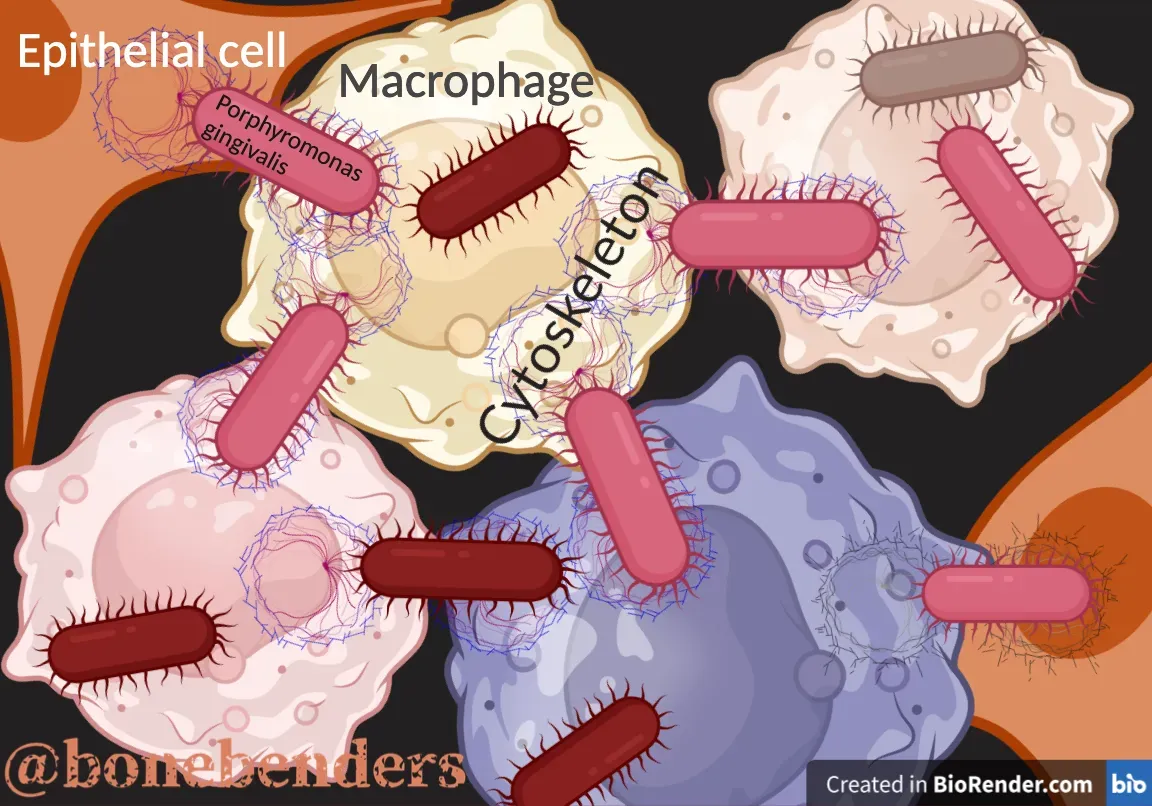
The physical invasion
The great microbial mass doesn’t limit itself to a chemical attack with toxins. P. gingivalis fights up close.
It physically penetrates the epithelial cells of the pocket, entering the cytoplasm. It moves from one cell to the next across bridges of the actin cytoskeleton. It thrives inside host cells — from epithelial cells to macrophages, the immune sentinels whose very job is to digest invaders.
The weapon is a hydrolytic enzyme that suppresses ATP-dependent apoptosis. Once inside the macrophage, P. gingivalis can disable its weapons and survive within it. But this is only the beginning.
The membrane receptors
The bacterium binds membrane integrin β1 through its major fimbriae. This activates FAK and paxillin, remodelling the actin cytoskeleton to open the door. Invasion requires actin polymerisation and microtubule formation. After 24 hours you can see a significant reassembly of the actin and tubulin filament network inside the host cells.
In essence, the bacterium borrows the contractile machinery of the host cell’s cytoplasm to move around inside it.
The outer membrane vesicles
P. gingivalis also uses outer membrane vesicles (OMVs), rich in gingipains and fimbriae, to facilitate cell invasion. These vesicles enter epithelial cells by endocytosis more efficiently than whole bacteria do. Once inside, they degrade epithelial proteins and ease bacterial adhesion.
Recent studies have identified more than 300 proteins in P. gingivalis OMVs — gingipains, fimbriae and peptidyl-arginine deiminase (PPAD) among them — confirming the role of these vesicles as vectors of virulence capable of reshaping the behaviour of oral fibroblasts.
A 2026 systematic review pooling 62 studies confirms the picture: the OMVs don’t stop at the periodontal microenvironment. They cross the circulation, reaching the brain, the heart, the liver. They induce ferroptosis in periodontal ligament stem cells and in bone mesenchymal cells. They modulate macrophages and dendritic cells. They are the most efficient vector P. gingivalis has.
That they actually travel — and it isn’t just an inference — was shown by Lv and colleagues in 2026, who tagged the OMVs with a fluorophore and tracked them in mice (BMC Oral Health). The distribution is time-dependent: at six hours the accumulation in liver and lung is sharp, and by twenty-four hours the lung signal has already faded. The detail that matters most: the vesicles are found in the cytoplasm of target cells in distant organs, not just in the intercellular matrix. They don’t merely arrive nearby — they get inside.
How it fools the immune system
Here the strategy turns subtle. P. gingivalis exploits the host protein CD47 to evade the immune response. CD47 is the “don’t eat me” signal — the same mechanism tumour cells use to escape immune surveillance. Switching CD47 on tells the white cell: “I’m one of yours, leave me alone.”
When CD47 is blocked with neutralising antibodies, P. gingivalis’s intracellular survival collapses sharply.
The protein thrombospondin-1 (TSP-1) partners with CD47 to repress the bactericidal activity of both macrophages and neutrophils. A double molecular shield.
Dismantling the immune defences
The gingipains don’t just facilitate invasion. They degrade key host immune proteins: LL-37, an antimicrobial peptide able to bind amyloid-beta and block its aggregation; apolipoprotein E; the antiviral interferons. This systematic demolition weakens innate immunity and sets the stage for persistent infection.
A 2026 study added a fine-grained piece: in macrophages, P. gingivalis suppresses the transcription of STAT1 — the central factor of the interferon-gamma pathway — cutting the expression of roughly 41% of the genes IFN-γ normally switches on. The mechanism needs direct contact and the Type IX Secretion System. Strains stripped of all three gingipains (Kgp, RgpA, RgpB) lose the ability to silence STAT1, and with it the ability to invade cells. Intracellular virulence and interferon sabotage are the same molecular programme.
It survives inside our own cells
P. gingivalis doesn’t just invade. It persists inside cells in a dormant state — not culturable in the lab, but alive and detectable. This is why periodontal infections are so hard to eradicate: the bacterium hides where antibiotics don’t reach efficiently.
The consequences reach beyond the mouth. P. gingivalis has been found in atherosclerotic tissue, in the brains of Alzheimer’s patients, and is associated with risks during pregnancy, gastric cancer and rheumatoid arthritis. A 2026 paper added colorectal cancer to the tally: the bacterium’s secreted factors — gingipains and hydrogen sulphide — drive epithelial-mesenchymal transition in intestinal cells, tuning the Wnt/β-catenin and Hippo-YAP pathways. And the liver? A 2026 study in the Journal of Nanobiotechnology linked periodontal severity to metabolic fatty liver disease (MASLD): the peptidyl-arginine deiminase carried by OMVs accumulates in the liver and suppresses the circadian regulator NPAS2, throttling fatty-acid oxidation. The line between oral infection and chronic systemic disease dissolves one piece at a time.
The mouth-gut-brain axis
Recent research has shown that oral P. gingivalis infection alters the gut microbiota, compromises the intestinal barrier and switches on systemic inflammatory responses. In particular, the bacterium disturbs tryptophan metabolism through the kynurenine pathway, producing neurotoxic metabolites such as 3-hydroxykynurenine that drive neuronal apoptosis. A route that connects the periodontal pocket to the brain by way of the gut.
Here too, the gingipains are the hinge. A paper published in Microbiome in 2026 showed that oral administration of P. gingivalis in mice induces gut dysbiosis and an expansion of Th17 cells — with a massive release of pro-inflammatory cytokines. Sequential knockouts of the gingipain genes progressively blunt the inflammatory response; when all three gingipains are absent, the inflammation eases significantly. The proteases aren’t accessory. They’re the engine.
The gingipains, moreover, can compromise the blood-brain barrier, letting virulence factors — outer membrane vesicles included — reach brain tissue directly, where they trigger neuroinflammation and promote the build-up of amyloid-beta and the hyperphosphorylation of tau.
A 2025 experiment in zebrafish larvae (Brain, Behavior, and Immunity) isolated the decisive variable. The wild-type W83 strain survives in the brain, activates the microglia and lights up the pro-inflammatory genes; the gingipain-null mutant is phagocytosed and cleared quickly, leaving no inflammatory trace. But the most instructive point is another: injecting the bacterium directly into the brain produces only a mild, transient inflammation, while it’s the systemic infection — the one starting from the periphery — that wrecks the blood-brain barrier. It isn’t the bacterium itself that punctures the brain. It’s the spread from the body that opens the way.
The fight against this bacterium is not a matter of hygiene. It’s a cellular war where the enemy uses molecular guerrilla tactics, hiding inside our own cells, subverting our defences and, from there, reaching distant organs along routes we are only beginning to understand. It’s the same logic that shapes how we treat severe periodontitis: break the chain of attack rather than chase its consequences.
And it all begins with something banal — colonisation. P. gingivalis is transmitted through the oral microbiome, and it isn’t the only pathobiont that has learned to get inside us.
Image created with BioRender.com.
FAQ
What is the role of Porphyromonas gingivalis in periodontal pockets?
Porphyromonas gingivalis colonises the depths of the periodontal pocket, where it multiplies aggressively by exploiting an anaerobic environment, driving the progression of periodontal disease.
How does P. gingivalis adapt to the pocket environment?
The bacterium thrives in a dark, oxygen-poor space, drawing energy from fermentation and anaerobic respiration, metabolising proteins and amino acids and releasing compounds such as fatty acids, gases and amines.
How does P. gingivalis invade host cells?
P. gingivalis physically penetrates epithelial cells by binding integrin β1 through its fimbriae, remodelling the actin cytoskeleton. It moves cell-to-cell across cytoskeletal bridges and uses gingipain-laden outer membrane vesicles to facilitate invasion.
How does it evade the immune system?
The bacterium exploits the host protein CD47 as a “don’t eat me” signal, working with thrombospondin-1 to suppress the bactericidal activity of macrophages and neutrophils. Its gingipains also degrade key immune proteins such as LL-37 and interferons.
Why is it so hard to eradicate P. gingivalis for good?
The bacterium persists inside cells in a dormant, non-culturable but still living state, hiding where antibiotics struggle to reach. This intracellular persistence also explains its links to systemic diseases such as Alzheimer’s.
References
- Amano A, et al. Molecular interactions of Porphyromonas gingivalis fimbriae with host proteins: kinetic analyses based on surface plasmon resonance. Cell Microbiol. 2002;4(6):331-338. doi:10.1046/j.1462-5822.2002.00192.x
- Yilmaz Ö, et al. Intercellular spreading of Porphyromonas gingivalis through host cell cytoskeleton. J Cell Biol. 2004;166(6):879-887. doi:10.1083/jcb.200406151
- CD47 and thrombospondin-1 contribute to immune evasion by Porphyromonas gingivalis. PNAS. 2024;121(47):e2405534121. doi:10.1073/pnas.2405534121 · PMID: 39536084
- Barron AE, et al. The dysregulation of innate immunity by Porphyromonas gingivalis in the aetiology of Alzheimer’s disease. J Intern Med. 2026;299(3). doi:10.1111/joim.70060 · PMID: 41424314
- Zhu H, et al. Porphyromonas gingivalis induces disturbance of kynurenine metabolism through the gut-brain axis. J Dent Res. 2025;104(4):439-448. doi:10.1177/00220345241303141
- Shawkatova I, et al. Alzheimer’s disease and Porphyromonas gingivalis: exploring the links. Life (Basel). 2025;15(1):96. doi:10.3390/life15010096 · PMID: 39860036
- Abe S, et al. Porphyromonas gingivalis suppresses interferon-gamma signaling in macrophages through a contact-dependent, gingipain-mediated mechanism. MicrobiologyOpen. 2026;15(2):e70290. doi:10.1002/mbo3.70290 · PMID: 41968448
- Li M, et al. Porphyromonas gingivalis induces intestinal inflammation through gingipain-dependent gut microbiome dysbiosis. Microbiome. 2026;14(1). doi:10.1186/s40168-026-02389-7 · PMID: 41923122
- Kazelnik M, et al. Porphyromonas gingivalis secreted factors drive epithelial-mesenchymal transition through gingipains and an H₂S-mediated bacterial defense system. Gut Microbes. 2026;18(1):2647532. doi:10.1080/19490976.2026.2647532 · PMID: 41873832
- Li Z, et al. Systematic review: Porphyromonas gingivalis outer membrane vesicles from pathogenesis to therapeutic implications. Int Dent J. 2026;76(3):109548. doi:10.1016/j.identj.2026.109548 · PMID: 41980468
- Widziolek M, et al. Porphyromonas gingivalis induces neuroinflammation in a gingipain-dependent manner in zebrafish larvae. Brain Behav Immun. 2025;132:106234. doi:10.1016/j.bbi.2025.106234 · PMID: 41423153
- Lv C, et al. Porphyromonas gingivalis-derived outer membrane vesicles travel from oral to distant organs via blood circulation in a time-dependent manner. BMC Oral Health. 2026;26(1). doi:10.1186/s12903-026-07979-3 · PMID: 41761257
- Liu Y, et al. Peptidylarginine deiminase in Porphyromonas gingivalis-derived outer membrane vesicles exacerbates metabolic dysfunction-associated steatotic liver disease through the NPAS2/CYP4A10 pathway. J Nanobiotechnology. 2026;24:342. doi:10.1186/s12951-026-04523-x · PMID: 42106850
FAQ
- What is the role of Porphyromonas gingivalis in periodontal pockets?
- Porphyromonas gingivalis colonises the depths of the periodontal pocket, where it multiplies aggressively by exploiting an anaerobic environment, driving the progression of periodontal disease.
- How does P. gingivalis adapt to the pocket environment?
- The bacterium thrives in a dark, oxygen-poor space, drawing energy from fermentation and anaerobic respiration, metabolising proteins and amino acids and releasing compounds such as fatty acids, gases and amines.
- How does P. gingivalis invade host cells?
- P. gingivalis physically penetrates epithelial cells by binding integrin β1 through its fimbriae, remodelling the actin cytoskeleton. It moves cell-to-cell across cytoskeletal bridges and uses gingipain-laden outer membrane vesicles to facilitate invasion.
- How does it evade the immune system?
- The bacterium exploits the host protein CD47 as a "don't eat me" signal, working with thrombospondin-1 to suppress the bactericidal activity of macrophages and neutrophils. Its gingipains also degrade key immune proteins such as LL-37 and interferons.
- Why is it so hard to eradicate P. gingivalis for good?
- The bacterium persists inside cells in a dormant, non-culturable but still living state, hiding where antibiotics struggle to reach. This intracellular persistence also explains its links to systemic diseases such as Alzheimer's.
References
- https://doi.org/10.1016/j.archoralbio.2025.106424
- https://doi.org/10.1046/j.1462-5822.2002.00192.x
- https://doi.org/10.1083/jcb.200406151
- https://doi.org/10.1111/joim.70060
- https://doi.org/10.1177/00220345241303141
- https://doi.org/10.3390/life15010096
- https://doi.org/10.1002/mbo3.70290
- https://doi.org/10.1186/s40168-026-02389-7
- https://doi.org/10.1016/j.identj.2026.109548
- https://doi.org/10.1080/19490976.2026.2647532
- https://doi.org/10.1016/j.bbi.2025.106234
- https://doi.org/10.1186/s12903-026-07979-3
- https://doi.org/10.1186/s12951-026-04523-x
Looking for a specialist?
Parodontologia a Frosinone →Diagnosi e trattamento della parodontite nello Studio Denti Più
Need a professional opinion?
Book an appointment at Dr. Bruschi's practice in Frosinone. First visit includes full diagnosis and personalised treatment plan.
Stay Updated
New articles on periodontology, implantology and oral surgery — delivered to your inbox.
Comments
Loading comments...
Leave a comment