Peri-implantitis with a buccal dehiscence: implantoplasty, xenograft and connective tissue graft in one session. Three years on
In brief — An anterior implant, one of six abutments under a metal-ceramic full-arch bridge, with peri-implantitis and titanium showing on the buccal side. In one session I treated every part of the defect: implantoplasty on the supracrestal portion, xenograft in the intraosseous component, and a connective tissue graft under a coronally advanced flap. At three years probing depth is 2 mm, there is no bleeding and the titanium is no longer visible. The full case report, with the photographs and a description of the technique, is on Zenodo as a preprint.
The patient was 60 at the time, with a metal-ceramic bridge on six implants placed by another clinician at least fifteen years earlier. He came in because of discomfort at one abutment, the upper left lateral incisor (22). When he lifted his lip you could see the polished implant collar and, under thin, oedematous mucosa, the start of the rough surface.
I measured a buccal probing depth of 6 mm, with bleeding on probing (BOP). There was a purulent infection. Keratinised tissue was under 2 mm and the mucosa was thin. The diagnosis was easy, frank peri-implantitis. The treatment choice less so.
One defect, two biologies: neighbouring, complementary, different
Periapical radiographs showed no mesial or distal bone loss. The lesion was entirely buccal, where lost bone projects over the implant body and disappears in two dimensions (periapical films). Had I seen only a radiograph, without visual inspection and the probe, I would have said the bone was there. The probe and the surface exposed above the crest made the diagnosis. But I saw the whole defect only when I raised the flap.
By the 2017 World Workshop definitions this is peri-implantitis [1,2], with an associated buccal soft-tissue dehiscence. The dehiscence measured 4 mm from the prosthetic margin: class II, subclass b in the Zucchelli classification [3]. That classification was built for single implants in the aesthetic zone, and I apply it here by analogy.
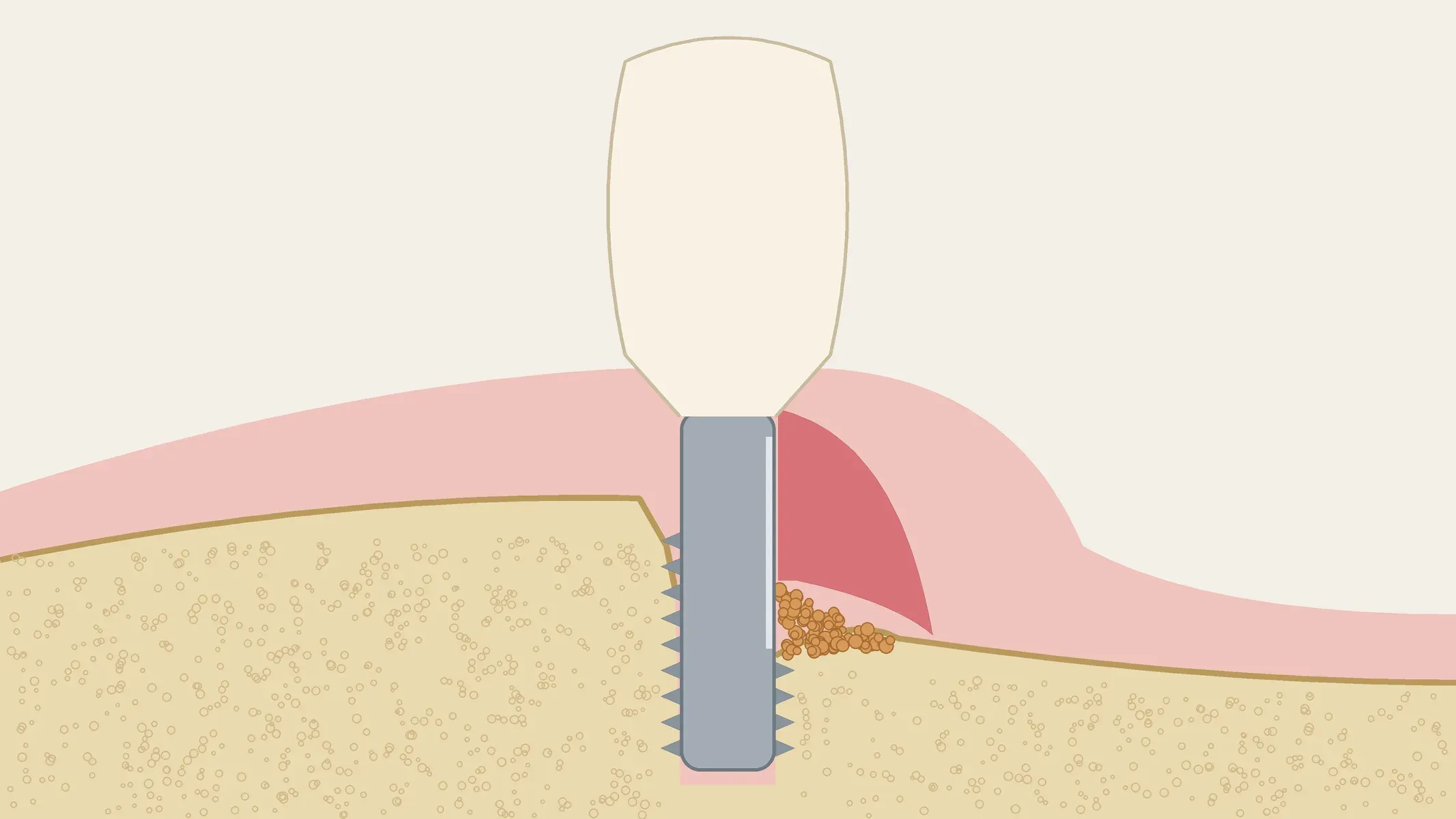
What decided everything was the shape of the defect. Inside the bone there was a partly contained crater. Above it, a buccal dehiscence with no walls, the titanium out of the crest and covered, where it was covered at all, by a millimetre of mucosa. Two problems with two different biological ceilings:
- in the contained part a bone substitute can fill the defect, and long-term data support it [4];
- in the wall-less part a graft has nothing to hold it, and re-osseointegration on a contaminated surface is unpredictable. There the goal becomes a smooth, non-retentive surface, then coverage.
At the time I did not have the TST technique, which today lets me avoid many implantoplasties. With a surface this rough, though, implantoplasty is probably still the most appropriate answer.
Why one technique was not enough
I discussed three alternatives with the patient and ruled all of them out.
Explantation meant dismantling a cemented arch for a defect that, at that stage, does well with surgery. Resective therapy alone controls the infection, as Romeo and colleagues showed at three years [5], but it increases recession, and in the anterior region it turns a biological success into an aesthetic failure. It leaves the patient a surface that is smooth, yes, but supragingival. Bone reconstruction alone makes no sense on the part of the defect that lacks a solid buccal wall.
That leaves the combined approach Schwarz described [6]: each component of the defect treated according to its own biology. I added the third piece, the soft tissue, in the same session. Soft tissue is usually managed later, once the peri-implantitis has resolved, or at non-infected sites; simultaneous soft-tissue volume augmentation during combined surgery has been reported only in small case series [7]. A thin phenotype matters for the implant’s health before it matters for looks. Thick, keratinised mucosa goes with less inflammation and a more stable margin [8], and that is what a patient needs who has to clean an abutment under a fixed arch every day. The histology of that soft-tissue cuff is in Zone A and Zone B.
The sequence
Before surgery the patient was treated by the dental hygienist: oral-hygiene instruction, supra- and submucosal instrumentation, air polishing and ultrasonic tips. The bridge was left in place, since it had anatomical emergence profiles and adequate space for cleaning. At re-evaluation two weeks later bleeding and suppuration persisted locally at implant 22, which confirmed the indication for surgery.
Prophylaxis was amoxicillin 2 g one hour before, the regimen the Cochrane review by Esposito supports for implant placement [9]. No prophylaxis trial exists for peri-implantitis surgery, so I applied it by extension. No antibiotic afterwards.
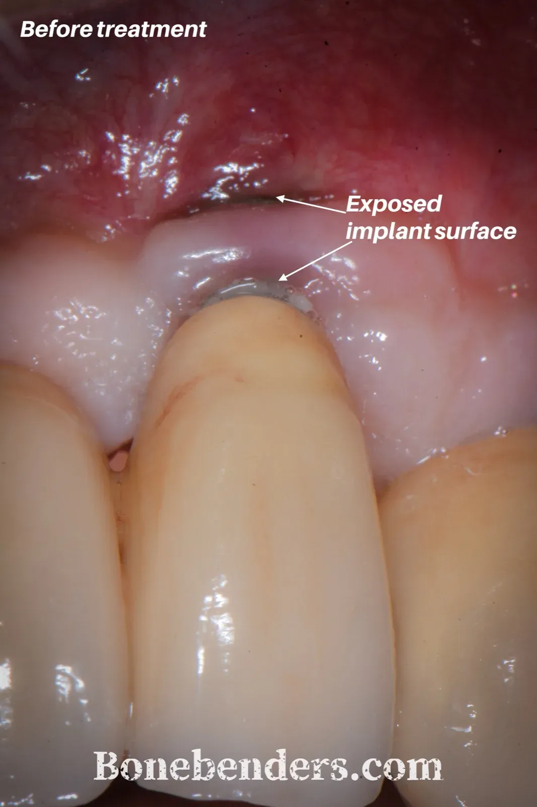
Figure A — Before treatment. The polished collar and the first part of the rough surface are exposed, under thin, oedematous, perforated mucosa.
Flap. Vertical releasing incisions and a buccal partial-thickness flap, leaving the periosteum on the bone focally, where the lesion had not already destroyed it and where the surface did not need exposing to remove the tissue soaked by the purulent infection.
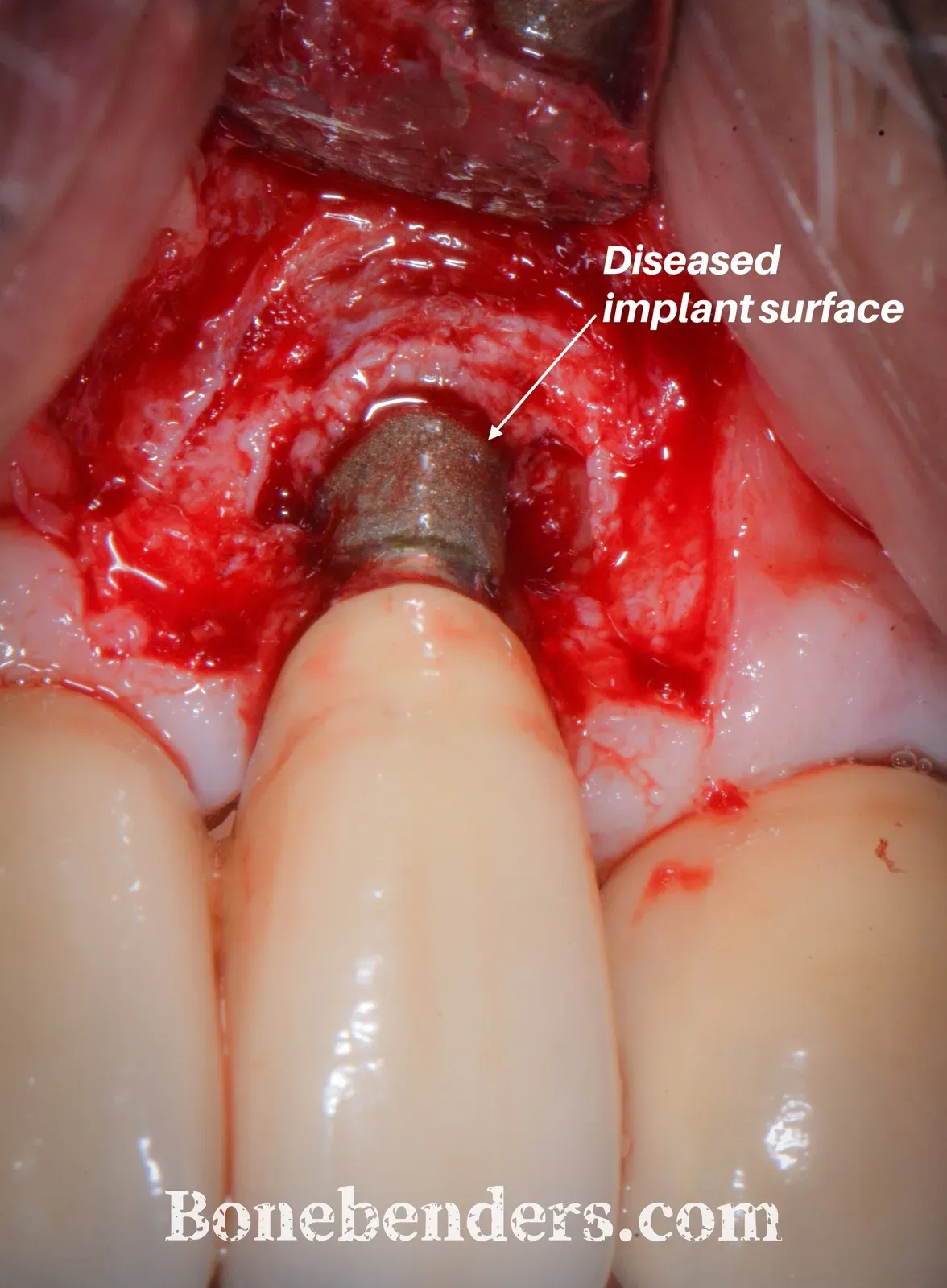
Figure B — The buccal defect with the flap open. The radiograph did not show it.
Debridement. Pus-soaked granulation tissue removed with titanium curettes and ultrasonic instruments under continuous saline irrigation.
Implantoplasty. Only on the supracrestal, dehisced portion: coarse then fine diamond burs, titanium brushes, silicone polishers, until the rough surface and any threads were gone and the surface was smooth and flush with the implant body. High-volume suction, copious irrigation and gauze to isolate the adjacent tissues. Then EDTA gel for 15 seconds and a rinse.
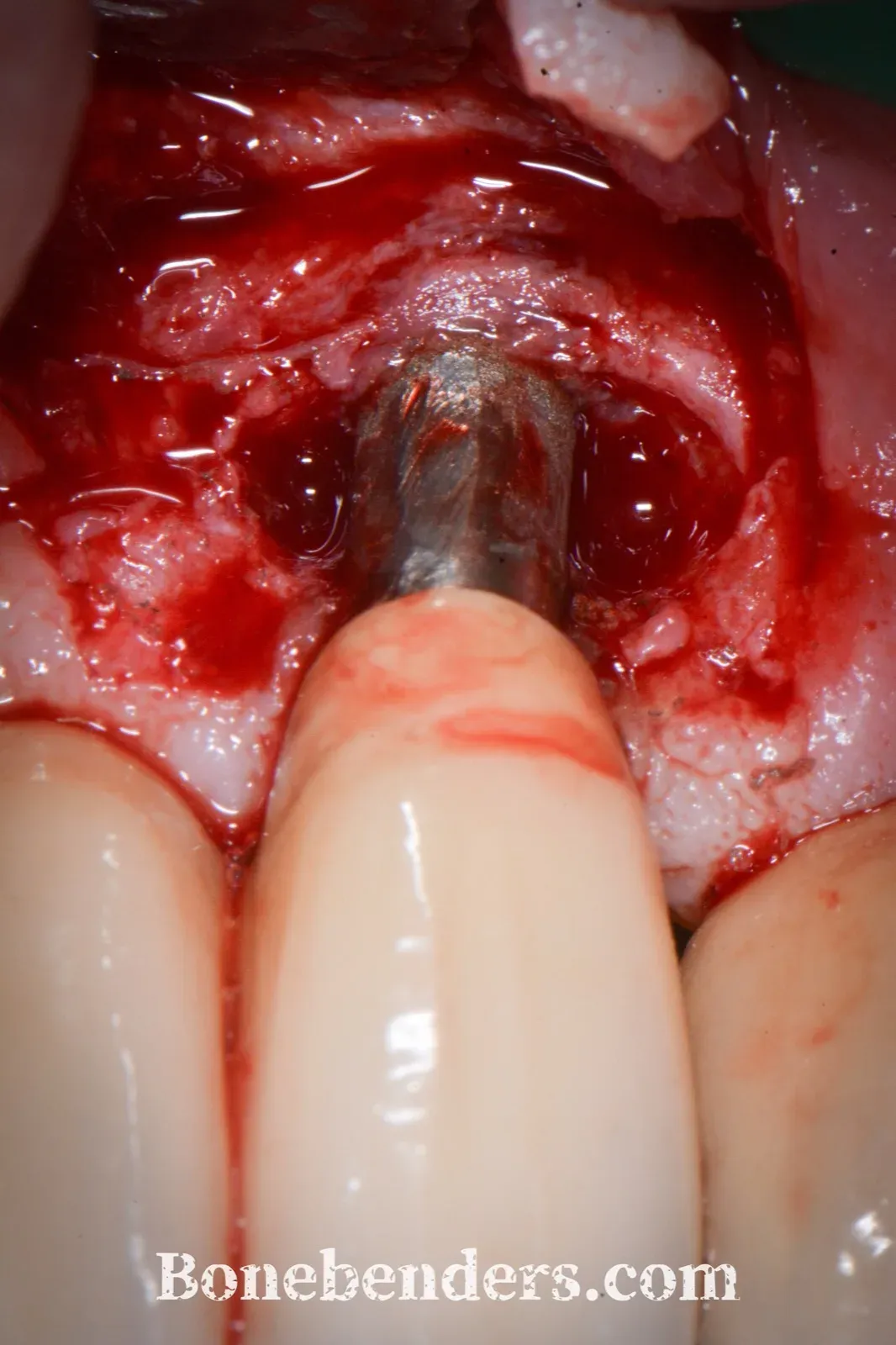
Figure C — After implantoplasty. Down in the crater the surface is still the original one.
Intraosseous component. Particulate xenograft hydrated with saline and condensed into the craters up to the level of the adjacent crest. No membrane over it, so that plasmatic perfusion reaches the connective tissue graft.
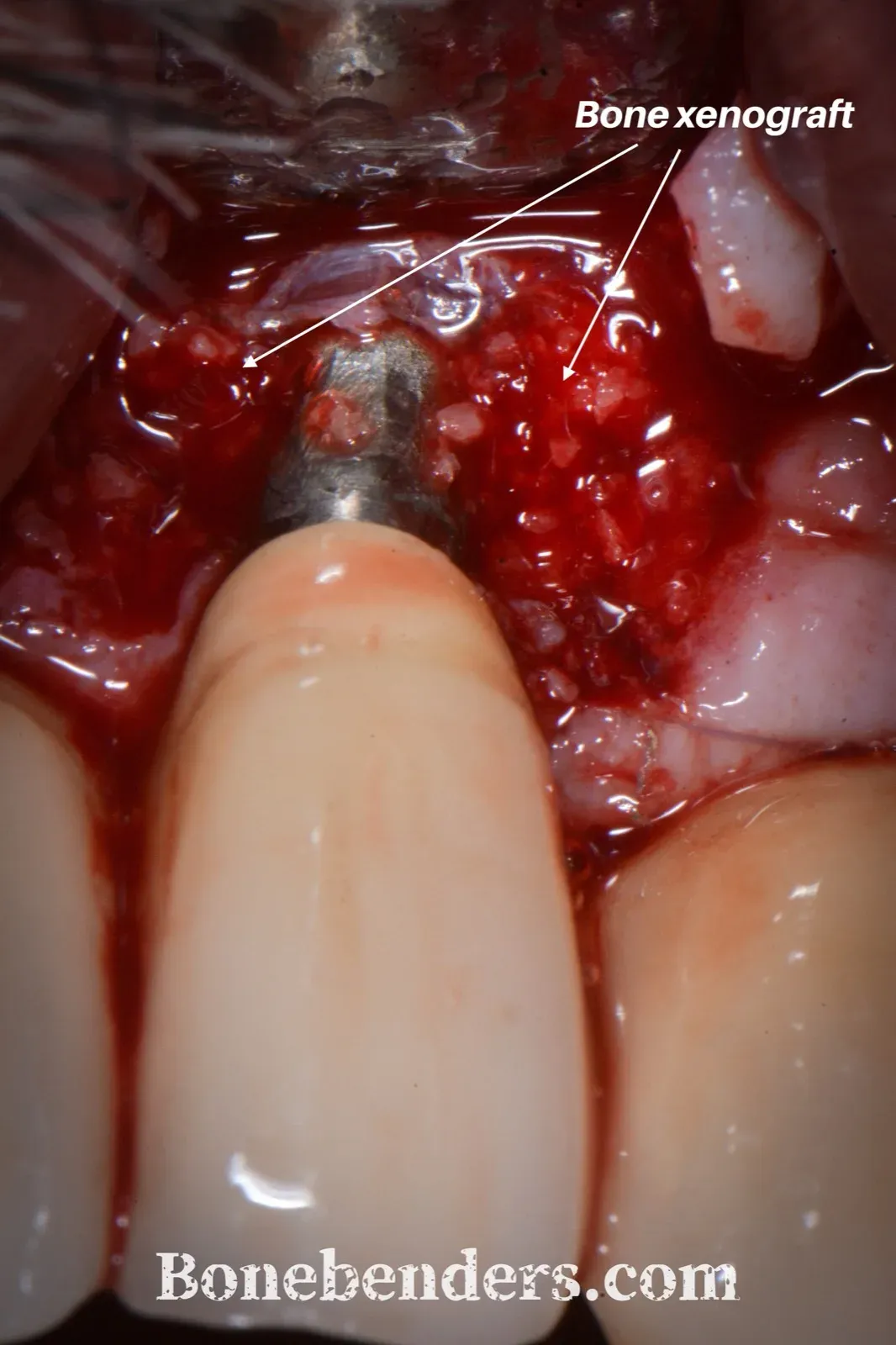
Figure D — Xenograft in the contained part of the defect.
Connective tissue graft. About 12 × 5 mm, harvested from the palate as a free gingival graft and then de-epithelialised. Placed over the defect to cover both the smoothed surface and the xenograft, and secured with 6-0 resorbable sutures. Compressed microfibrillar collagen haemostat on the donor site.
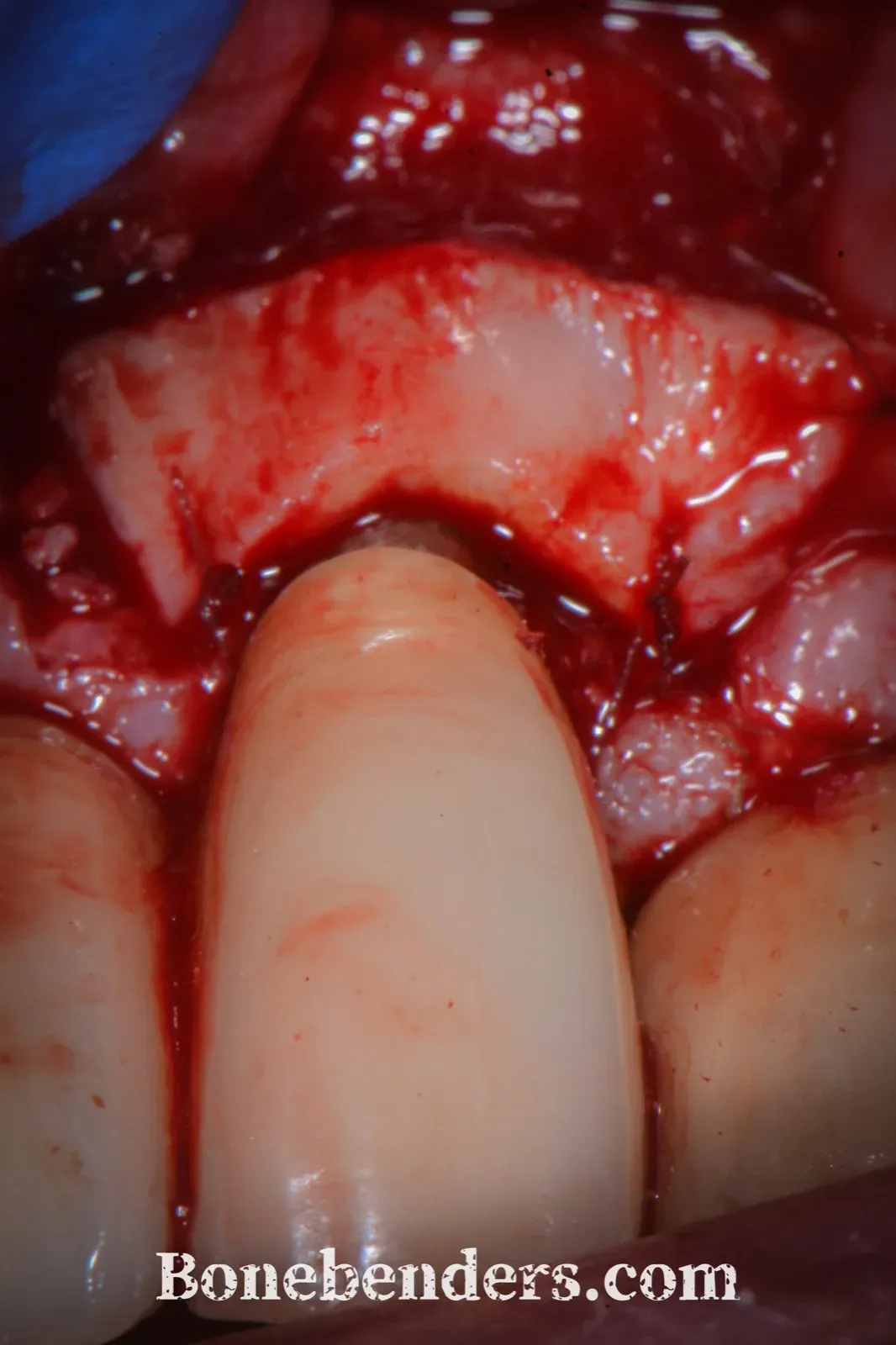
Figure E — One graft covers the smoothed titanium and the bone graft together.
Closure. Coronally advanced flap over the graft, tension-free after periosteal release, sutured with 6-0 resorbable sutures for complete primary coverage.
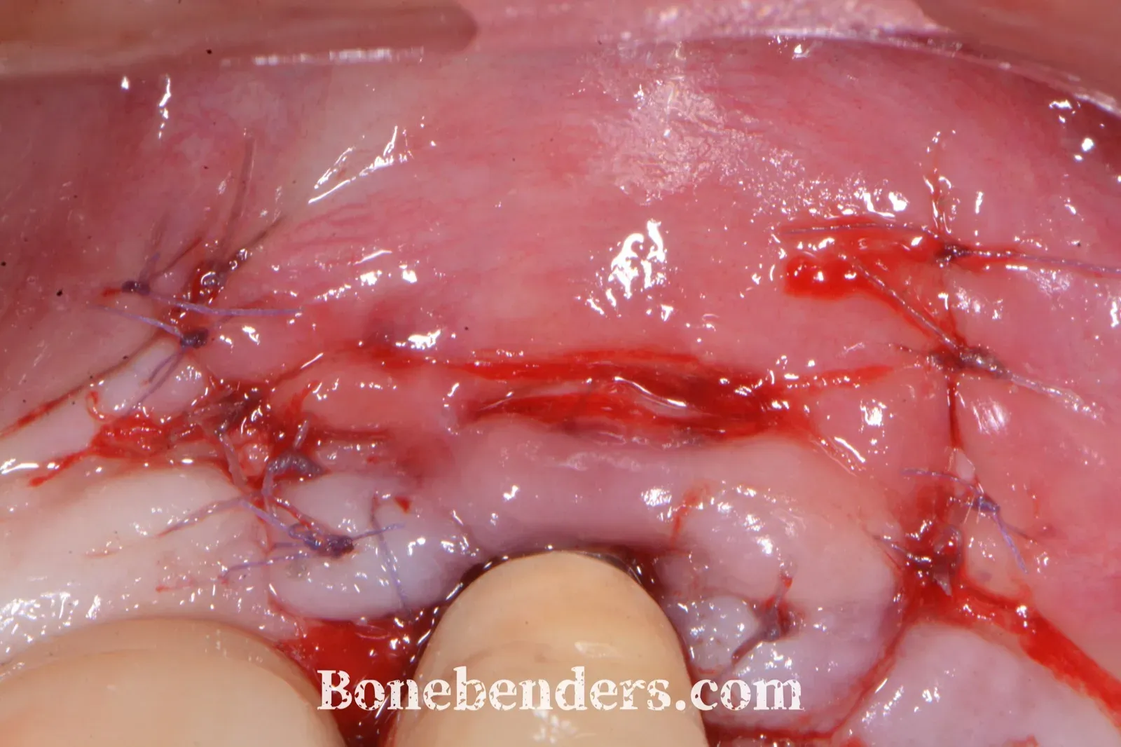
Figure F — Primary coverage of the graft.
Ibuprofen twice daily, soft diet, no brushing at the site for a week. Sutures out at 15 days. Uneventful healing, palate included.
Three years later
Reviews at 1, 2, 4 and 12 weeks, then at 6, 12, 24 and 36 months, with supportive peri-implant care every three months. The patient’s compliance was excellent; without it, none of what follows would mean much.
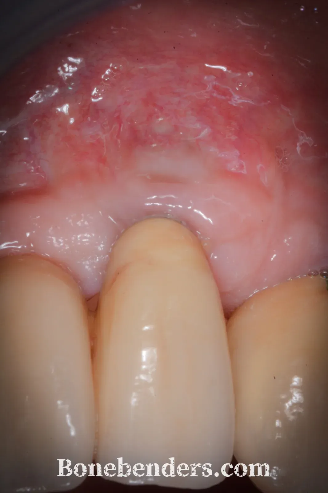
Figure G — At three years. Pink, healthy, firm mucosa with no oedema. No titanium in sight.
| Parameter | Baseline | 3 years |
|---|---|---|
| Deepest probing depth | 6 mm | 2 mm |
| Bleeding on probing | yes | no |
| Suppuration | yes | no |
| Buccal dehiscence height | 4 mm | absent |
| Patient-reported aesthetic satisfaction (VAS 0–10) | 2 | 8 |
By the composite criterion of resolution (no bleeding or suppuration on probing, probing depth up to 5 mm, no further bone loss) the case is resolved [10]. I did not re-measure mucosal thickness or keratinised tissue width. The gain is visibly evident.
In the case report I recorded his words at three years: “I sought the doctor’s consultation because of persistent pain at a dental implant that had been placed by another doctor many years earlier. The doctor explained that my implant was in bad shape and needed deep cleaning and a graft procedure. The procedure was free of pain, and there was hardly any swelling in the days following the surgery. After 30 days my implant was back to a perfect shape, without pain or metal showing — and it still is, after many years.”
The limits of implantoplasty
Three limitations, essentially.
The implant wall. Smoothing removes structure from the titanium. In laboratory testing implantoplasty did not change the fracture resistance of standard-diameter external-connection implants [11], but it significantly reduced the elastic limit of internal-connection implants, especially at 3–3.5 mm [12]. This patient’s implant measured 3.8 mm, make and system unknown. That is the low end of standard diameter.
The particles. The procedure scatters metallic debris into the tissues. Titanium particles are found in peri-implant tissues and have been linked to inflammatory responses, but a causal role in peri-implantitis progression has not been shown [13]. Isolation, suction and irrigation reduce dispersion, but they do not eliminate it. I have written about titanium as a material in tribocorrosion; here the issue is different, because the surgical procedure generates the debris.
The decontaminant. No agent has proved better than the others. Baima and colleagues pooled the decontamination protocols used during surgery and found comparable clinical outcomes [14]. The EDTA gel used here does not explain the result. The TST technique is a different matter; this case predates it.
What this case does not show
It is one case. No second, blinded examiner; measurements taken by me, who operated and followed the patient from start to finish. Probing depth depends on probe angulation and on soft-tissue thickness. There is no quantitative radiographic outcome, because the defect was buccal and cannot be measured in two dimensions. Three years is short in the life of an implant, and recurrence after peri-implantitis surgery is not rare over longer horizons.
A good outcome in one patient shows the sequence is feasible, but not necessarily that it works better than another. Implantoplasty, xenograft and connective tissue graft are three different procedures, but in this case they overlap, and there is no way to know what share of the final result each one contributed.
If you have a case like this
Before you choose the technique, look at the shape of the defect and split it. The contained part can be grafted. The wall-less part gets smoothed and covered. Mucosal thickness decides whether the patient will manage to keep it clean and maintain it over time.
If the radiograph is clean and the probe sinks only on the buccal side, trust the probe.
For a different approach to decontamination, in surgical treatment of peri-implantitis with the TST technique Dr De Nale regenerates a crater-shaped defect without implantoplasty.
The full case report — Bruschi E. Combined resective–reconstructive and mucogingival management of peri-implantitis with a buccal soft-tissue dehiscence at an anterior implant supporting a full-arch restoration: a 3-year case report. Zenodo; 2026. doi:10.5281/zenodo.23351880. Manuscript with CARE checklist, 30 references and the seven original photographs at full capture resolution.
References
- Berglundh T, Armitage G, Araujo MG, Avila-Ortiz G, Blanco J, Camargo PM, et al. Peri-implant diseases and conditions: consensus report of workgroup 4 of the 2017 World Workshop on the Classification of Periodontal and Peri-Implant Diseases and Conditions. J Clin Periodontol. 2018;45 Suppl 20:S286-91. doi:10.1111/jcpe.12957 · PMID: 29926491
- Renvert S, Persson GR, Pirih FQ, Camargo PM. Peri-implant health, peri-implant mucositis, and peri-implantitis: case definitions and diagnostic considerations. J Clin Periodontol. 2018;45 Suppl 20:S278-85. doi:10.1111/jcpe.12956 · PMID: 29926496
- Zucchelli G, Tavelli L, Stefanini M, Barootchi S, Mazzotti C, Gori G, et al. Classification of facial peri-implant soft tissue dehiscence/deficiencies at single implant sites in the esthetic zone. J Periodontol. 2019;90(10):1116-24. doi:10.1002/JPER.18-0616 · PMID: 31087334
- Roccuzzo M, Fierravanti L, Pittoni D, Dalmasso P, Roccuzzo A. Implant survival after surgical treatment of peri-implantitis lesions by means of deproteinized bovine bone mineral with 10% collagen: 10-year results from a prospective study. Clin Oral Implants Res. 2020;31(8):768-76. doi:10.1111/clr.13628 · PMID: 32559322
- Romeo E, Ghisolfi M, Murgolo N, Chiapasco M, Lops D, Vogel G. Therapy of peri-implantitis with resective surgery. A 3-year clinical trial on rough screw-shaped oral implants. Part I: clinical outcome. Clin Oral Implants Res. 2005;16(1):9-18. doi:10.1111/j.1600-0501.2004.01084.x · PMID: 15642026
- Schwarz F, John G, Mainusch S, Sahm N, Becker J. Combined surgical therapy of peri-implantitis evaluating two methods of surface debridement and decontamination. A two-year clinical follow up report. J Clin Periodontol. 2012;39(8):789-97. doi:10.1111/j.1600-051X.2012.01867.x · PMID: 22639800
- Schwarz F, Sahm N, Becker J. Combined surgical therapy of advanced peri-implantitis lesions with concomitant soft tissue volume augmentation. A case series. Clin Oral Implants Res. 2014;25(1):132-6. doi:10.1111/clr.12103 · PMID: 23350647
- Tavelli L, Barootchi S, Avila-Ortiz G, Urban IA, Giannobile WV, Wang HL. Peri-implant soft tissue phenotype modification and its impact on peri-implant health: a systematic review and network meta-analysis. J Periodontol. 2021;92(1):21-44. doi:10.1002/JPER.19-0716 · PMID: 32710810
- Esposito M, Grusovin MG, Worthington HV. Interventions for replacing missing teeth: antibiotics at dental implant placement to prevent complications. Cochrane Database Syst Rev. 2013;(7):CD004152. doi:10.1002/14651858.CD004152.pub4 · PMID: 23904048
- Herrera D, Berglundh T, Schwarz F, Chapple I, Jepsen S, Sculean A, et al. Prevention and treatment of peri-implant diseases—The EFP S3 level clinical practice guideline. J Clin Periodontol. 2023;50 Suppl 26:4-76. doi:10.1111/jcpe.13823 · PMID: 37271498
- Costa-Berenguer X, García-García M, Sánchez-Torres A, Sanz-Alonso M, Figueiredo R, Valmaseda-Castellón E. Effect of implantoplasty on fracture resistance and surface roughness of standard diameter dental implants. Clin Oral Implants Res. 2018;29(1):46-54. doi:10.1111/clr.13037 · PMID: 28736922
- Diéguez-Pereira M, Chávarri-Prado D, Viteri-Agustín I, Montalban-Vadillo O, Pérez-Pevida E, Brizuela-Velasco A. Effect of implantoplasty on the elastic limit of dental implants of different diameters. Int J Implant Dent. 2021;7(1):88. doi:10.1186/s40729-021-00363-6 · PMID: 34426894
- Suárez-López Del Amo F, Garaicoa-Pazmiño C, Fretwurst T, Castilho RM, Squarize CH. Dental implants-associated release of titanium particles: a systematic review. Clin Oral Implants Res. 2018;29(11):1085-100. doi:10.1111/clr.13372 · PMID: 30280418
- Baima G, Citterio F, Romandini M, Romano F, Mariani GM, Buduneli N, et al. Surface decontamination protocols for surgical treatment of peri-implantitis: a systematic review with meta-analysis. Clin Oral Implants Res. 2022;33(11):1069-86. doi:10.1111/clr.13992 · PMID: 36017594
- Bruschi E. Combined resective–reconstructive and mucogingival management of peri-implantitis with a buccal soft-tissue dehiscence at an anterior implant supporting a full-arch restoration: a 3-year case report. Zenodo; 2026. doi:10.5281/zenodo.23351880
Cover: original Bonebenders diagram.
FAQ
Why not regenerate the whole defect with bone graft?
Doesn't implantoplasty weaken the implant?
Wouldn't it have been simpler to remove the implant?
Doesn't grafting connective tissue in the same session put the bone graft at risk?
How much does the decontaminant matter?
Cite this article
Ready for theses, slides and papers. If you cite it on a web page, make the URL a link so readers can reach the source.
Bruschi E. Peri-implantitis with a buccal dehiscence: implantoplasty, xenograft and connective tissue graft in one session. Three years on [Internet]. Frosinone (Italy): Bonebenders; 2026 [cited 2026 Oct 14]. Available from: https://bonebenders.com/en/blog/peri-implantitis-implantoplasty-connective-tissue-graft-3-years/ Bruschi, E. (2026, October 14). Peri-implantitis with a buccal dehiscence: implantoplasty, xenograft and connective tissue graft in one session. Three years on. Bonebenders. https://bonebenders.com/en/blog/peri-implantitis-implantoplasty-connective-tissue-graft-3-years/
@misc{bruschi2026peri,
  author       = {Bruschi, Ernesto},
  title        = {Peri-implantitis with a buccal dehiscence: implantoplasty, xenograft and connective tissue graft in one session. Three years on},
  year         = {2026},
  month        = oct,
  howpublished = {Bonebenders},
  url          = {https://bonebenders.com/en/blog/peri-implantitis-implantoplasty-connective-tissue-graft-3-years/},
  note         = {Accessed 2026-10-14}
} Go deeper
Espansione crestale (split crest) →Allargare la cresta senza trapianto — tecnica ERE e bonebending
Need a professional opinion?
Book an appointment at Dr. Bruschi's practice in Frosinone. First visit includes full diagnosis and personalised treatment plan.
Stai valutando un impianto dentale?
Ti hanno detto che non hai abbastanza osso, o ti hanno proposto un’arcata fissa? Ho scritto una guida di dieci pagine: le strade possibili, gli esami che servono e le dieci domande da fare prima di firmare.
Ricevi la guidaStay Updated
New articles on periodontology, implantology and oral surgery — delivered to your inbox.
Comments
Loading comments...
Leave a comment