Buried Alive: The Osteoblast's Three Fates
Leggi in Italiano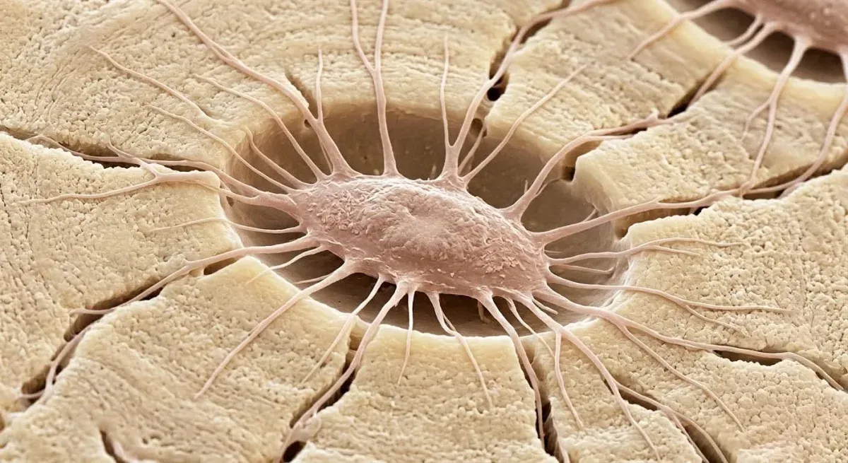
In brief — The osteoblast builds bone by laying down the matrix. Once the work is done, three fates await it: apoptosis, which claims most of them; the quiet of a lining cell resting on the surface; or burial in the matrix it has just made — buried alive. That last osteoblast becomes an osteocyte, the longest-lived bone cell — it lives for decades — and turns from builder into director of remodeling. For anyone who operates on bone, the point is this: you are not working a material, but inside a network of living cells that are already reading what you do.
There is a moment in bone that is fascinating and terrible at once. The osteoblast has finished. It has laid down the matrix, layer on layer, and now it stands still, surrounded by what it has built. It does not yet know what will happen to it. Or perhaps it does, if we consider it within the collective identity of the holobiont.
No bone cell is born an osteocyte. Each one was first an osteoblast that, at some point, stopped building. This is the story of that cell, hidden in the depths of the bone.
First the osteoid, then the bone
The osteoblast is the cell that builds. It sits on the surface of the bone, in orderly rows, and lays down osteoid. An organic matrix, still soft, made mostly of collagen. A support scaffold. Then the mineral arrives, settles inside that scaffold, and the osteoid hardens — it gradually becomes real bone.
It is work of constant renewal. Bone is not static, it is unmade and remade for a lifetime, and the osteoblast is the worker on this great bio-construction site. In 2006, Franz-Odendaal and colleagues described what can happen to this worker when the work slows. In some cases the osteoblast itself ends up embedded in the mineralized matrix it has just produced. The cell that raises the wall can stay inside the wall itself.
It is only one of the possible endings. When deposition stops, three roads open in front of the osteoblast that has just finished its work.
Three roads
When it has finished, the osteoblast does not have a single possible future. It has three.
The first is death. For most of these cells the fate is apoptosis. A programmed death, orderly, with no inflammation around it. Clean. The same procedure the body uses to clear out diseased cells, like those of cancer. Manolagas, in a review from 2000 that remains a landmark, puts it plainly: the life of bone rests on the birth and death of its cells. They are born, they work, they go, and this turnover holds the tissue in balance — it is the weave of bone remodeling, the constant unmaking and remaking. Most of the cells that build your bone will never see it finished. Each laid down its share of matrix, and then went dark.
The second is rest, followed by a different life. Some osteoblasts neither die nor are buried. They flatten, stretch out over the surface, and stay there, quiet. They are the base cells of the tissue. They cover the bone, keep watch over it. But they are still osteoblasts, and at a signal they can turn active again and go back to laying matrix. A reserve parked on the threshold. Cheap to keep, and worth a great deal when you can call it back at need.
The third fate is the strangest of them all.
Buried alive. An end or a new beginning?
The third osteoblast stays inside.
While its neighbors go on laying down matrix, it slows. It produces less, then stops. And the others’ matrix reaches it: surrounds it, covers it, closes it inside the new bone. Franz-Odendaal, Hall and Witten made it clear: the burial is passive. No one pushes the osteoblast into the wall. It simply stops outrunning it. It slows, and the matrix catches up.
Buried alive, they called it. Walled in alive, in the house it built.
But is this an end, or a new beginning?
It looks like an end. The body flattens, the cell stays shut in a small non-calcified niche of the bone matrix, and it will never lay down bone again. And yet the longest life in bone begins right there. That buried osteoblast becomes an osteocyte. Dozens of processes reach out from the flattened body, running through the finest canaliculi in search of the others like it, buried around. A node is born, then a network.
Here the scale changes. Osteocytes are 90-95% of all the cells in adult bone, and the longest-lived: they last for decades, far longer than the osteoblasts that gave rise to them (Bonewald, 2011). And from inside, they are not idle. They sense load, strain, the fluid that moves when bone bends, and on those signals they decide where to resorb and where to deposit, commanding osteoclasts and osteoblasts (Dallas, Prideaux and Bonewald, 2013). The builder, buried alive, has become the director of the skeleton.
How that network is built — the canaliculi, the dialogue, the way it reads forces — is another story, and I told it in the osteocyte connectome. Here we focus on the shift: from worker to director.
What changes, for the operator
When we expand a ridge, when we move a cortical plate, when we graft bone, the temptation is to treat it as a material: hard, inert, to be shaped like a piece of wood. Under the instrument, though, there is a population of living cells — osteocytes walled into the matrix we are cutting, moving, compressing. They are already reading what we do. They feel the pressure of the instrument, the cut, every flex that passes through the matrix. Bone is an inhabited tissue, and it answers back.
To move a cortical plate, then, is to hand that network a new piece of information, and to wait for its reply in the weeks that follow. Anyone who operates with measure keeps this in mind: calibrated movements, respect for biology’s own timing, the awareness that every maneuver is a message read by an entity that was already there. In the end it is that cell, buried alive, that weighs on how much of the bone we have moved will consolidate, and on how well its osteointegration will hold over the years.
References
- Franz-Odendaal TA, Hall BK, Witten PE. Buried alive: how osteoblasts become osteocytes. Dev Dyn. 2006;235(1):176-90. doi:10.1002/dvdy.20603. PMID: 16258960.
- Manolagas SC. Birth and death of bone cells: basic regulatory mechanisms and implications for the pathogenesis and treatment of osteoporosis. Endocr Rev. 2000;21(2):115-37. doi:10.1210/edrv.21.2.0395. PMID: 10782361.
- Bonewald LF. The amazing osteocyte. J Bone Miner Res. 2011;26(2):229-38. doi:10.1002/jbmr.320. PMID: 21254230.
- Dallas SL, Prideaux M, Bonewald LF. The osteocyte: an endocrine cell … and more. Endocr Rev. 2013;34(5):658-90. doi:10.1210/er.2012-1026. PMID: 23612223.
FAQ
What is the difference between an osteoblast and an osteocyte?
What does it mean that the osteoblast is “buried alive”?
Why does osteoblast biology matter to bone surgery?
Cite this article
Ready for theses, slides and papers. If you cite it on a web page, make the URL a link so readers can reach the source.
Bruschi E. Buried Alive: The Osteoblast's Three Fates [Internet]. Frosinone (Italy): Bonebenders; 2026 [cited 2026 Oct 25]. Available from: https://bonebenders.com/en/blog/buried-alive-osteoblast-three-fates/ Bruschi, E. (2026, September 6). Buried Alive: The Osteoblast's Three Fates. Bonebenders. https://bonebenders.com/en/blog/buried-alive-osteoblast-three-fates/
@misc{bruschi2026buried,
  author       = {Bruschi, Ernesto},
  title        = {Buried Alive: The Osteoblast's Three Fates},
  year         = {2026},
  month        = sep,
  howpublished = {Bonebenders},
  url          = {https://bonebenders.com/en/blog/buried-alive-osteoblast-three-fates/},
  note         = {Accessed 2026-10-25}
} Go deeper
Espansione crestale (split crest) →Allargare la cresta senza trapianto — tecnica ERE e bonebending
Need a professional opinion?
Book an appointment at Dr. Bruschi's practice in Frosinone. First visit includes full diagnosis and personalised treatment plan.
Stai valutando un impianto dentale?
Ti hanno detto che non hai abbastanza osso, o ti hanno proposto un’arcata fissa? Ho scritto una guida di dieci pagine: le strade possibili, gli esami che servono e le dieci domande da fare prima di firmare.
Ricevi la guidaStay Updated
New articles on periodontology, implantology and oral surgery — delivered to your inbox.
Comments
Loading comments...
Leave a comment