Alveolar bone: what it is, why it is lost, and how you rebuild volume
Leggi in Italiano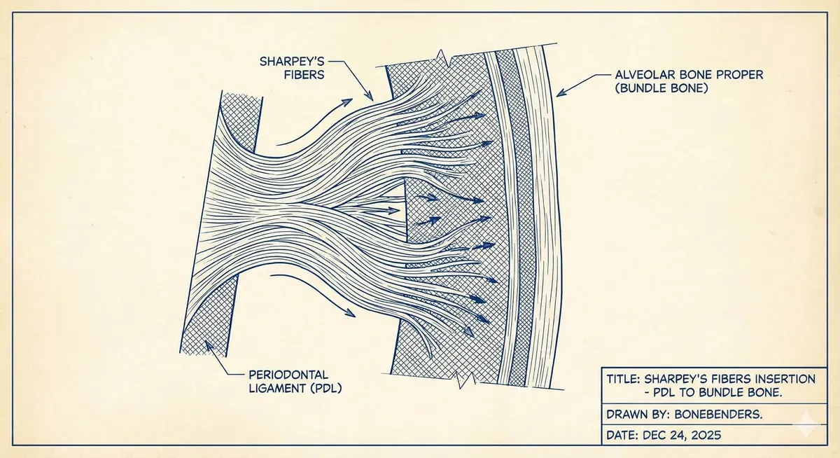
In brief — Alveolar bone proper depends on the tooth and the ligament. After extraction it resorbs in a predictable pattern. Implants do not rebuild it. Buccal wall thickness, extraction trauma, and — when needed — ridge preservation set the plan.
Pull a tooth and, within weeks, the ridge changes shape. That is physiology of alveolar bone proper — bundle bone, the lamina dura you read on radiographs — not a surgical mishap.
Tan and colleagues (Clinical Oral Implants Research, 2012) pooled human data on post-extraction change: within six months horizontal loss runs 29–63% (about 3.8 mm on average) and vertical loss 11–22% (about 1.2 mm). Those ranges explain why an implant plan drawn on the pre-extraction volume often fails in the mouth.
Alveolar process versus alveolar bone proper
The alveolar process is the tooth-bearing part of the jaws. Three layers: outer cortices (buccal and palatal/lingual), intervening spongiosa, and alveolar bone proper lining the socket and anchoring the periodontal ligament.
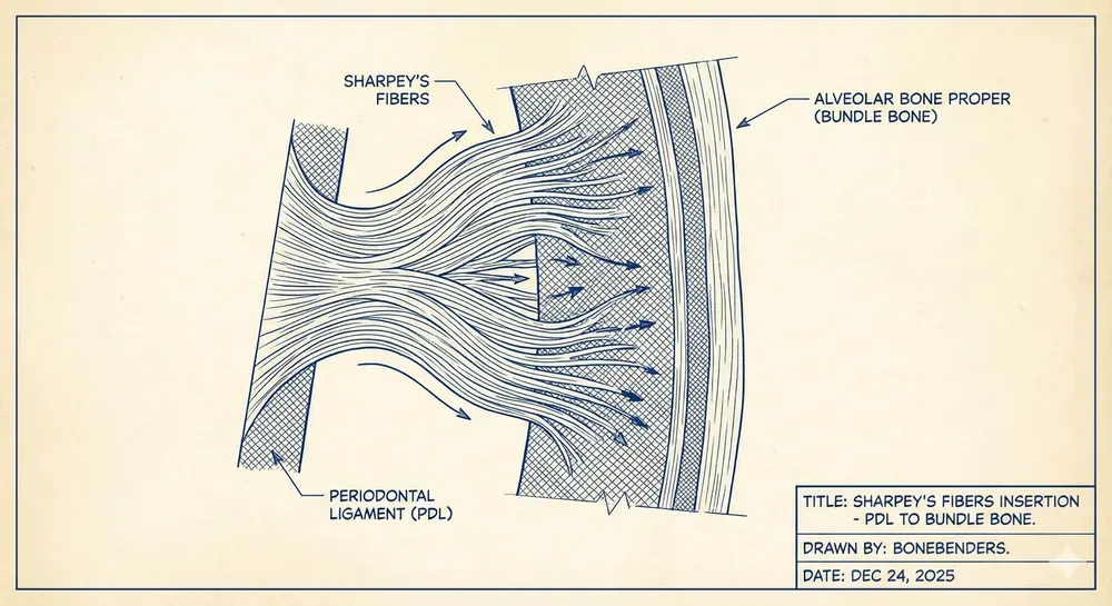
Alveolar bone proper is a 0.2–0.4 mm sheet of circumferential lamellae without a mature Haversian system. Sharpey fibers insert there — hence bundle bone. Blood arrives from the ligament through Volkmann canals. Remove the tooth and that circuit shuts down. That biology underpins the socket-shield proof-of-principle by Hürzeler and colleagues (Journal of Clinical Periodontology, 2010): leave a buccal root fragment so ligament and wall volume can stay.
On parallel periapicals the same tissue reads as lamina dura: a continuous radiopaque line 0.2–0.3 mm along the root. Two-dimensional films read that line well. Buccal thickness, they do not. For that you need CBCT — when diagnosis or implant planning asks for it, not as automatic routine.
Why resorption follows extraction
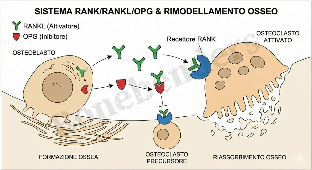
Bundle-bone loss follows functional dependence on the tooth and ligament. RANKL/RANK/OPG steers osteoclastogenesis; without periodontal stimulus, osteoclasts clear the lamina that lost its blood supply.
Araújo and Lindhe (Journal of Clinical Periodontology, 2005) and Cardaropoli and colleagues (2003) mapped socket healing in the dog. In the first 1–2 weeks bundle bone is replaced by woven bone; TRAP-positive cells already sit on the surface by day three. Between weeks 2 and 8, outer cortical resorption continues — a step the original authors still called poorly understood. By four weeks the socket is filled with woven bone; lamellar bone and marrow follow over the next months.
Buccal wall thickness sets the volume
Chappuis and colleagues (Journal of Dental Research, 2013) followed 39 patients with serial CBCT in the esthetic zone. At eight weeks, mid-buccal vertical median loss was 5.2 mm (48% of height); horizontal only 0.3 mm. Proximally, median vertical loss was 0.5 mm. Buccal thickness ≤1 mm drove the split: thin-wall phenotypes lost 7.5 mm vertically (62%), thick-wall only 1.1 mm (9%). Their 2017 Periodontology 2000 review restated why those millimeters matter clinically.
Monje, Roccuzzo, Buser and Wang (Clinical Oral Implants Research, 2023) revisited buccal bone wall thickness in a systematic review: thin walls after implant placement show larger dimensional change and vertical bone loss, with mucosal recession risk. One millimeter of buccal cortex outweighs a millimeter of surface marketing.
Ickroth and colleagues (Clinical Oral Implants Research, 2024) meta-analyzed six RCTs (222 single implants in the esthetic zone) comparing immediate and early placement. With an intact buccal wall and low-risk patients, esthetics and survival did not diverge significantly. Certainty stays low — most trials carried high or unclear bias — yet the clinical point is clear: wall integrity matters more than the “immediate versus early” calendar.
Ridge preservation: what newer data add
Atieh and colleagues’ Cochrane review (2021) included 16 RCTs (426 adults, 524 sites). Xenografts cut width loss (MD about −1.2 mm) and height loss (MD about −1.4 mm) versus extraction alone, but certainty is very low, and need for extra augmentation was not statistically different in that synthesis. Read it as a caution on evidence quality, not a dismissal of grafting.
Wei and colleagues (Journal of Evidence-Based Dental Practice, 2025) focused on molars: 7 studies, 237 sockets in 236 patients. Versus spontaneous healing, ridge preservation reduced horizontal change (MD 2.21 mm) and mid-buccal vertical change (MD 1.34 mm), and lowered the risk of needing augmentation at implant placement (RR 0.41; 95% CI 0.26–0.65). At molars the clinical gain is easier to see.
Cecchinato and colleagues (Journal of Periodontology, 2026) randomized 42 patients who needed two adjacent extractions: DBBM-C plus collagen membrane versus spontaneous healing. At six months horizontal reduction was 57.7% in controls and 23.0% in grafted sites; buccal vertical loss 3.0 mm versus 1.5 mm. Neighboring sockets shrink harder — grafting changes those numbers clearly.
Lu and colleagues (International Journal of Implant Dentistry, 2025) meta-analyzed 27 studies (1307 implants) on socket shield versus conventional immediate placement in the esthetic zone: less horizontal and vertical buccal bone loss, better pink esthetic scores, comparable implant success. Technique-sensitive, protocols still poorly standardized — useful when anatomy and experience allow, not a universal shortcut.
Surgical trauma and planning
Flap elevation increases resorption versus a flapless approach that keeps periosteal supply. Pre-existing periodontal or periapical inflammation worsens healing; clear infection first when you can. For timing after single-rooted extractions, see also immediate implants and bone defects and the practical notes on the first 48 hours after extraction.
When volume is already gone, site classification — for example the Wang A1–C3 alveolar bone map — steers preservation, expansion, or grafting. An expander inside an atrophic socket is one graftless path when anatomy fits. Place the implant where stable bone and coherent soft tissue remain, not where the tooth sat on last year’s radiograph.
FAQ
Why does bundle bone (lamina dura) matter for implant surgery?
Can you limit post-extraction ridge loss?
How do alveolar process and alveolar bone proper differ?
What drives post-extraction resorption biologically?
What does lamina dura imaging tell you before implants?
References
- Tan WL, Wong TL, Wong MC, et al. A systematic review of post-extractional alveolar hard and soft tissue dimensional changes in humans. Clin Oral Implants Res. 2012;23 Suppl 5:1-21. doi:10.1111/j.1600-0501.2011.02375.x · PMID: 22211303
- Chappuis V, Engel O, Reyes M, et al. Ridge alterations post-extraction in the esthetic zone: a 3D analysis with CBCT. J Dent Res. 2013;92(12 Suppl):195S-201S. doi:10.1177/0022034513506713 · PMID: 24158340
- Chappuis V, Araújo MG, Buser D. Clinical relevance of dimensional bone and soft tissue alterations post-extraction in esthetic sites. Periodontol 2000. 2017;73(1):73-83. doi:10.1111/prd.12167 · PMID: 28000281
- Araújo MG, Lindhe J. Dimensional ridge alterations following tooth extraction. An experimental study in the dog. J Clin Periodontol. 2005;32(2):212-218. doi:10.1111/j.1600-051x.2005.00642.x · PMID: 15691354
- Cardaropoli G, Araújo M, Lindhe J. Dynamics of bone tissue formation in tooth extraction sites. An experimental study in dogs. J Clin Periodontol. 2003;30(9):809-818. doi:10.1034/j.1600-051x.2003.00366.x · PMID: 12956657
- Hürzeler MB, Zuhr O, Schupbach P, et al. The socket-shield technique: a proof-of-principle report. J Clin Periodontol. 2010;37(9):855-862. doi:10.1111/j.1600-051x.2010.01595.x · PMID: 20712701
- Atieh MA, Alsabeeha NH, Payne AG, et al. Interventions for replacing missing teeth: alveolar ridge preservation techniques for dental implant site development. Cochrane Database Syst Rev. 2021;4(4):CD010176. doi:10.1002/14651858.cd010176.pub3 · PMID: 33899930
- Monje A, Roccuzzo A, Buser D, et al. Influence of buccal bone wall thickness on the peri-implant hard and soft tissue dimensional changes: A systematic review. Clin Oral Implants Res. 2023;34 Suppl 26:8-27. doi:10.1111/clr.14177 · PMID: 37750522
- Wei YP, Han ZY, Hu WJ, et al. ALVEOLAR RIDGE PRESERVATION AT MOLAR EXTRACTION SITES: A SYSTEMATIC REVIEW AND META-ANALYSIS. J Evid Based Dent Pract. 2025;25(1):102074. doi:10.1016/j.jebdp.2024.102074 · PMID: 39947777
- Cecchinato D, Corrà E, Bressan E, et al. Biomaterial used to counteract ridge reduction following the removal of adjacent teeth: A randomized controlled multicenter study. J Periodontol. 2026;97(7):1481-1490. doi:10.1002/jper.70084 · PMID: 41870328 · free full text
- Ickroth A, Seyssens L, Christiaens V, et al. Immediate versus early implant placement for single tooth replacement in the aesthetic area: A systematic review and meta-analysis. Clin Oral Implants Res. 2024;35(6):585-597. doi:10.1111/clr.14261 · PMID: 38558205
- Lu W, Du S, Su J, et al. Clinical efficacy of socket shield technique compared to conventional immediate implant placement in the aesthetic zone: a meta-analysis. Int J Implant Dent. 2025;11(1):72. doi:10.1186/s40729-025-00657-z · PMID: 41417433 · free full text
- Monje A, Roccuzzo A, Buser D, et al. Influence of buccal bone wall thickness on the peri-implant hard and soft tissue dimensional changes: A systematic review. Clin Oral Implants Res. 2023;34 Suppl 26:8-27. doi:10.1111/clr.14177 · PMID: 37750522
- Wei YP, Han ZY, Hu WJ, et al. ALVEOLAR RIDGE PRESERVATION AT MOLAR EXTRACTION SITES: A SYSTEMATIC REVIEW AND META-ANALYSIS. J Evid Based Dent Pract. 2025;25(1):102074. doi:10.1016/j.jebdp.2024.102074 · PMID: 39947777
- Cecchinato D, Corrà E, Bressan E, et al. Biomaterial used to counteract ridge reduction following the removal of adjacent teeth: A randomized controlled multicenter study. J Periodontol. 2026;97(7):1481-1490. doi:10.1002/jper.70084 · PMID: 41870328 · free full text
- Ickroth A, Seyssens L, Christiaens V, et al. Immediate versus early implant placement for single tooth replacement in the aesthetic area: A systematic review and meta-analysis. Clin Oral Implants Res. 2024;35(6):585-597. doi:10.1111/clr.14261 · PMID: 38558205
Cite this article
Ready for theses, slides and papers. If you cite it on a web page, make the URL a link so readers can reach the source.
Bruschi E. Alveolar bone: what it is, why it is lost, and how you rebuild volume [Internet]. Frosinone (Italy): Bonebenders; 2026 [updated 2026 Aug 20; cited 2026 Oct 22]. Available from: https://bonebenders.com/en/blog/alveolar-bone-why-it-is-lost-and-how-it-is-rebuilt/ Bruschi, E. (2026, January 9). Alveolar bone: what it is, why it is lost, and how you rebuild volume. Bonebenders. https://bonebenders.com/en/blog/alveolar-bone-why-it-is-lost-and-how-it-is-rebuilt/
@misc{bruschi2026alveolar,
  author       = {Bruschi, Ernesto},
  title        = {Alveolar bone: what it is, why it is lost, and how you rebuild volume},
  year         = {2026},
  month        = jan,
  howpublished = {Bonebenders},
  url          = {https://bonebenders.com/en/blog/alveolar-bone-why-it-is-lost-and-how-it-is-rebuilt/},
  note         = {Accessed 2026-10-22}
} Go deeper
Espansione crestale (split crest) →Allargare la cresta senza trapianto — tecnica ERE e bonebending
Need a professional opinion?
Book an appointment at Dr. Bruschi's practice in Frosinone. First visit includes full diagnosis and personalised treatment plan.
Stai valutando un impianto dentale?
Ti hanno detto che non hai abbastanza osso, o ti hanno proposto un’arcata fissa? Ho scritto una guida di dieci pagine: le strade possibili, gli esami che servono e le dieci domande da fare prima di firmare.
Ricevi la guidaStay Updated
New articles on periodontology, implantology and oral surgery — delivered to your inbox.
Comments
Loading comments...
Leave a comment