The Expander Inside the Socket: Immediate Implant in Atrophic Bone
Leggi in Italiano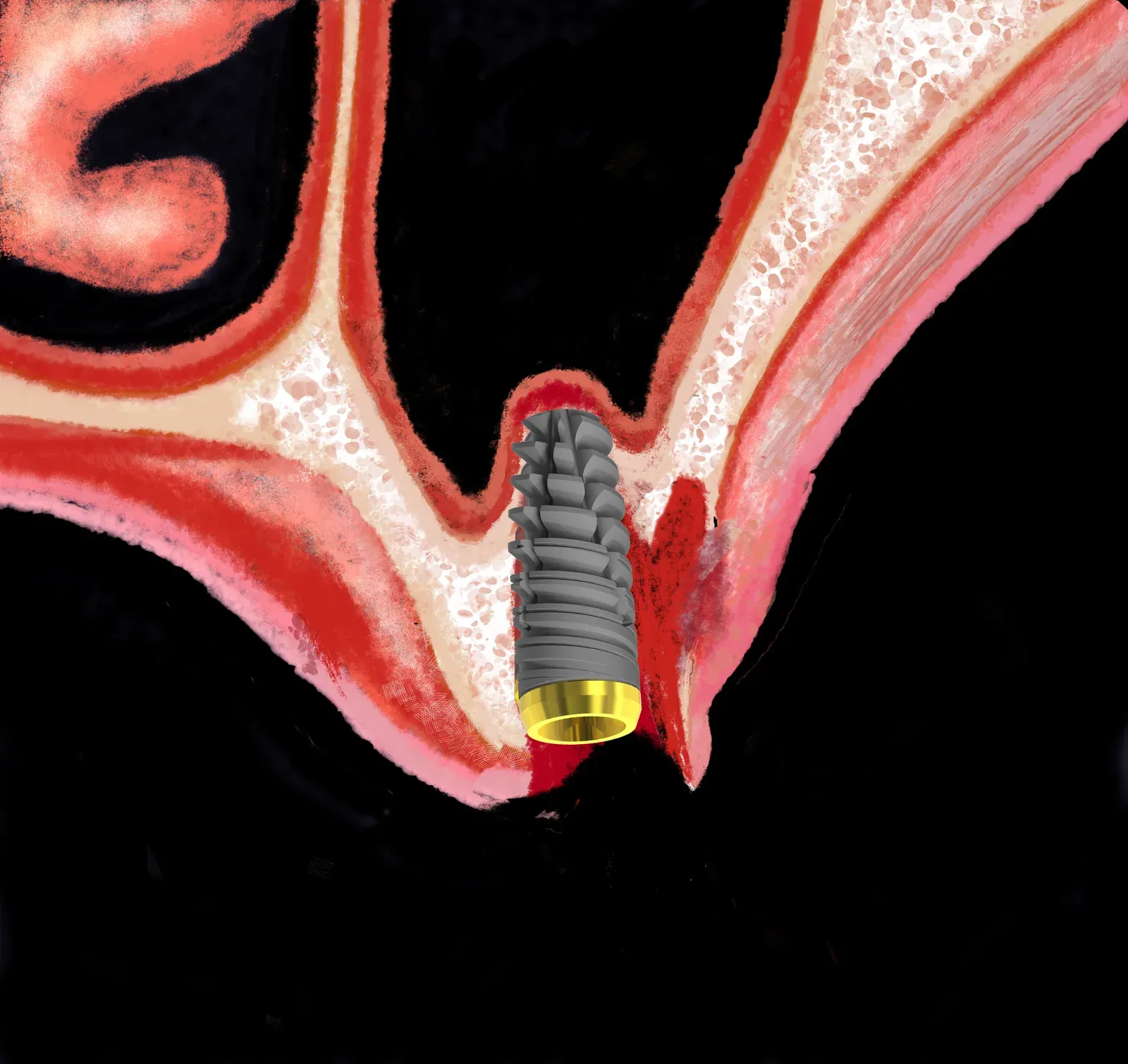
In brief — Expanders work inside a fresh socket and in severely atrophic sites. The socket is a natural guide; walls are compressed rather than removed; the implant finds stability in residual bone, without drills that take more of it away.
The socket is not an obstacle. It is a guide.
At the same time, it can be the wrong guide. You have to reshape that path for your own use. Keep what helps; discard what leads you off course. Expanders are the tools for that job. Unique instruments, with real handling and directionality.
The socket shows both the wrong axis and the right one. Once you find the right direction, the goal is to lock one or more anchorage points along it. Expanders are built for that precision — the kind of precision that decides primary stability in a post-extraction site.
That shift in perspective is where everything else begins. And if you have any sense, you will see the chance to do something rare for your patients — and for yourself.
The classic territory of ridge expansion
Edentulous ridge expansion (ERE) was designed for the healed edentulous ridge: narrow, with two corticals and expandable marrow. A controlled cut, lateral displacement, implant insertion. The internal cancellous bone regenerates. This is the canonical territory, codified in 1994 by Scipioni and Bruschi.¹
But the healed ridge is not the only place where an expander can work. The post-extraction site takes the technique past its classic borders — and that is where the difference from a conventional drill becomes decisive.
The post-extraction site
The tooth must be extracted. Surrounding bone is scarce. Resorption is chronic, periodontitis advanced; what remains are long thin roots in little bony structure.
The conventional choice is to wait: extract, allow 3-4 months, reassess. The problem is that socket preservation does not always deliver what is promised, especially when chasing the usual “innovations” of the moment. Waiting also throws away the osteoregenerative capacity of the periodontal ligament (PDL) cells that remain after extraction.
The alternative is to extract and place the implant in the same session, using the socket as a “guide that is not a guide” for the expander.
It is a guide when the correct implant position matches the axis of the extracted root.
More often it is a non-guide that is still useful. Root position rarely coincides with the ideal implant site. In that case the implant emerges from the socket, but the apex diverges in search of the right position and of maximum stability in solid native bone.
Biology is on your side: the fresh socket is rich in clot, platelet-derived growth factors, mesenchymal progenitor cells, and the PDL cells mentioned above. This is the most metabolically active window. Waiting means losing it.
Surgical sequence
Atraumatic extraction. Preserving the alveolar walls is the primary objective. Periotomy with thin instruments, low-force luxators, no aggressive forceps. The more intact the walls, the more material the expander has to work with.
Socket decontamination. Thorough curettage of granulation tissue, generous irrigation. If the tooth was in active septic condition — abscess, fistula, osteitis — the procedure must be reassessed: an infected socket is not a reliable starting point.
Sounding and axis definition. The smallest expander travels through the canal and reads the resistance of the walls. The future implant axis does not necessarily coincide with the axis of the extracted root: divergence is often necessary to follow available bone.
Progressive expansion. From smaller to larger diameters, slowly. Tactile feedback tells you when the wall is yielding elastically and when rigidity signals the limit. Never force beyond that signal.
Implant insertion. Insertion torque is the reality test. Above 30-35 Ncm, proceed with the prosthetic timeline; below that threshold, reassess loading timing. In very soft type IV bone, deferred loading is often the more prudent choice.²
Gap management. A space between the implant surface and the socket walls is nearly always present. Up to 2 mm, the clot holds. Beyond that, a low-density particulate graft is considered. Membrane and primary closure depend on morphology: no universal rules apply.
CBCT is mandatory
None of these decisions can be improvised. CBCT is the only tool that measures alveolar wall thickness in all dimensions, checks the distance from the maxillary sinus or inferior alveolar nerve, and allows the implant axis to be planned. Personally, I prefer the intraoperative scan.
A panoramic radiograph is not enough. Sagittal and coronal sections at the site of interest are the mandatory starting point.
When it works and when it does not
It works when alveolar walls are present and continuous — even thin —, the site is free of active infection, the correct implant axis is compatible with local anatomy, and the operator knows the tactile feedback of expanders and knows where to stop. The operator must also know the resistance pillars of the maxilla, such as the supramaxillary arch.
It can also work when the vestibular wall is fenestrated or completely resorbed, provided local anatomy still allows a correct axis and solid apical mechanical anchorage in basal bone, or close to it.
References
- Scipioni A, Bruschi GB, Calesini G. The edentulous ridge expansion technique: a five-year study. Int J Periodontics Restorative Dent 1994;14(5):451-9. PubMed
- Crespi R, Bruschi GB, Gastaldi G, Capparé P, Gherlone EF. Immediate loaded implants in split-crest procedure. Clin Implant Dent Relat Res 2015;17 Suppl 2:e692-8. PubMed
- Bruschi GB, Crespi R, Capparé P, Bravi F, Grande N, Gherlone E, Gastaldi G. Radiographic evaluation of crestal bone level in split-crest and immediate implant placement: minimum 5-year follow-up. Int J Oral Maxillofac Implants 2017;32(1):114-120. PubMed
- Scipioni A, Bruschi GB, Calesini G, Bruschi E, De Martino C. Bone regeneration in the edentulous ridge expansion technique: histologic and ultrastructural study of 20 clinical cases. Int J Periodontics Restorative Dent 1999;19(3):269-77. PubMed
FAQ
Can an implant be placed immediately after extraction in atrophic bone?
Why use an expander instead of a drill when bone is very scarce?
What is the socket and what role does it play in this technique?
How thick must the alveolar wall be to use an expander?
How is the gap between implant and socket walls managed?
What are the risks of immediate post-extraction implant placement with expanders?
Cite this article
Ready for theses, slides and papers. If you cite it on a web page, make the URL a link so readers can reach the source.
Bruschi E. The Expander Inside the Socket: Immediate Implant in Atrophic Bone [Internet]. Frosinone (Italy): Bonebenders; 2026 [cited 2026 Oct 22]. Available from: https://bonebenders.com/en/blog/socket-expanders-post-extraction-bone-atrophy/ Bruschi, E. (2026, April 18). The Expander Inside the Socket: Immediate Implant in Atrophic Bone. Bonebenders. https://bonebenders.com/en/blog/socket-expanders-post-extraction-bone-atrophy/
@misc{bruschi2026socket,
  author       = {Bruschi, Ernesto},
  title        = {The Expander Inside the Socket: Immediate Implant in Atrophic Bone},
  year         = {2026},
  month        = apr,
  howpublished = {Bonebenders},
  url          = {https://bonebenders.com/en/blog/socket-expanders-post-extraction-bone-atrophy/},
  note         = {Accessed 2026-10-22}
} Go deeper
Espansione crestale (split crest) →Allargare la cresta senza trapianto — tecnica ERE e bonebending
Need a professional opinion?
Book an appointment at Dr. Bruschi's practice in Frosinone. First visit includes full diagnosis and personalised treatment plan.
Stai valutando un impianto dentale?
Ti hanno detto che non hai abbastanza osso, o ti hanno proposto un’arcata fissa? Ho scritto una guida di dieci pagine: le strade possibili, gli esami che servono e le dieci domande da fare prima di firmare.
Ricevi la guidaStay Updated
New articles on periodontology, implantology and oral surgery — delivered to your inbox.
Comments
Loading comments...
Leave a comment