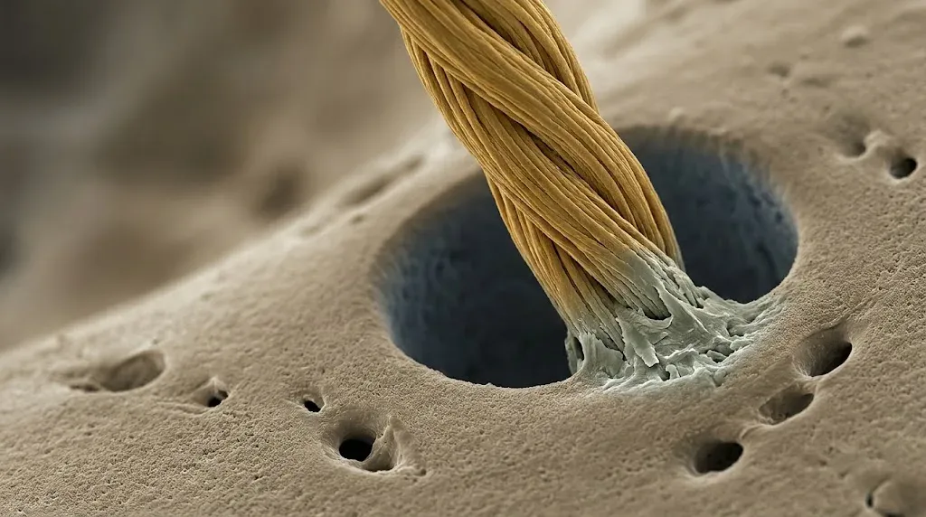
In brief — Periosteal Sharpey’s fibers are rich in type III collagen, 5-25 µm thick and poorly mineralized. Osteoclasts leave them alone, so they outlast bone remodeling. Some cross the entire cortex and fuse with the endosteum. Jean E. Aaron calls this continuum PSE (Periosteum, Sharpey’s fiber, Endosteum) and proposes it as the histological substrate of Frost’s “mechanostat.” The network shrinks when estrogen falls, expands with exercise and fragments with age. Anyone raising a full-thickness flap should reckon with consequences that reach all the way to the marrow.
Sharpey’s fibers are a second-year topic. Perforating fibers: they anchor the periosteum to the cortex and the periodontal ligament to alveolar bone. They also turn up across the whole skeleton, wherever tendons, ligaments and periosteum meet bone. In 2012 Jean E. Aaron, of the Bone Structural Biology Laboratory in Leeds, published a fine review in Frontiers in Endocrinology titled Periosteal Sharpey’s fibers: a novel bone matrix regulatory system? The hypothesis is that these fibers help govern bone homeostasis and remodeling.
The Scottish anatomist William Sharpey named them perforating fibres in the mid-nineteenth century, because they pass straight through the lamellae, piercing them. They had been described long before, in Rome, by Domenico Gagliardi: his 1689 Anatome ossium speaks of claviculi, “nails that pierce the lamellae of bone and hold them together.”
What is new is the idea that they form a regulatory system for bone metabolism.
What Aaron’s review says
Immunohistochemistry changed the picture. Once type III collagen could be labeled, those pale, barely visible fibers turned into an obvious surface network.
The collagen is type III — in bone, where you would expect type I — and with it come type VI collagen, elastin and tenascin. Thickness: 5 to 25 µm. Above all, they are poorly mineralized, and that is the most interesting fact of the lot. With so little mineral the osteoclast essentially cannot see them, so they survive bone remodeling.
They enter the cortex at different angles. Oblique fibers are the most numerous and typical of young bone. Vertical ones cross the full thickness and reach in between the trabeculae. Horizontal, superficial ones become more common with age. The insertion pattern is multiaxial and the composition complex, far more than a purely anchoring job would need. Aaron builds the whole hypothesis on that mismatch.
Some of these fibers keep going. They cross the entire cortex, reach the endosteum and fuse with a very thin layer of type III-rich osteoid — under 2 µm — that lines every resting bone surface.
This is where Aaron introduces the concept: PSE, Periosteum – Sharpey’s Fibre – Endosteum. A biofunctional continuum. The two membranes of bone, outer and inner, which we have always studied as separate entities, would be one structure, stitched through the cortex by Sharpey’s fibers. A sac that wraps bone from outside and inside at once.
The sac moves, remodels and takes bone with it
Here the review moves from anatomy to physiology, the part that concerns us most directly.
Estrogen. Ovariectomized rats, a model of menopause. The type III-rich proximal domain contracts and the fibers disconnect. This happens before bone matrix is lost, as if the reorganization of the fiber system were steering bone structure.
Mechanical loading. Rats with free access to a running wheel. The subperiosteal fiber domain widens by 50% at midshaft and fibers labeled for type III collagen increase by 15%. Around them the cortex grows (+18% proximal width, +7% midshaft area) and trabecular volume rises 25%, with new interconnections. The fiber network changes, and bone structure follows.
Age. Pig mandible, 1-year-olds against 3-year-olds. The periosteum thins (180 to 129 µm), the fibers shorten dramatically (226 to 55 µm) and fragment, leaving mostly horizontal, superficial ones. Calcified microparticles about 1 µm across start to appear, in irregular clusters or wrapped around the bundles.
Disease. Human proximal femur. In osteoporosis the fiber network is atrophic; in osteoarthritis, hypertrophic.
Aaron’s conclusion is elegant, and it should be taken for what it is: a hypothesis. The Sharpey’s fiber network could be the histological substrate of Frost’s mechanostat, the physical place where bone decides whether to grow or shrink.
Why should we care?
We operate mostly on bone of intramembranous origin — the bone where Sharpey’s fibers are most at home.
And above all, we strip the periosteum every time we raise a full-thickness flap. Aaron’s work suggests we may be tearing away the outer end of a mechanobiological system that reaches the marrow, and the evidence on flap design in ridge splitting shows that the gesture has measurable effects on the bone left behind.
A few considerations follow. They seem reasonable to me, though they remain considerations rather than certainties.
The flap has a biological cost we don’t measure. We measure volume, millimeters, atrophy. We don’t measure fibrous architecture — and in this model, that architecture decides whether the site remodels well or badly.
The periosteum as a membrane stands on firmer ground than we thought. Ma and colleagues (Scientific Reports, 2023) compared periosteal coverage with a collagen membrane in a canine buccal dehiscence model. The periosteum group gained more vertical bone, formed more new bone and mineralized faster. It is an animal model, not a final verdict, but it points the same way and deserves a place in any discussion of GBR and autologous grafts.
And the implant? An implant has no ligament. No perforating fiber enters it. At the implant site the PSE continuum is broken by definition. That is part of what osseointegration means: we have traded a network of responsive fibers for a rigid interface. It works very well. It is also a different physiology, and the implant lives as a guest inside a complex biological system.
A short history of Sharpey’s fibers
For a hundred and fifty years the literature on Sharpey’s fibers was almost entirely dental. The fibers were the anchor of the periodontal ligament in bundle bone, and that was it. Aaron turns the telescope around, taking what we learned in the periodontium and extending it to the whole skeleton.
Bone, all bone, seems to behave like a periodontium. It responds to what its fibers do.
The review closes by proposing that type III-rich fibers may be as essential for holding functioning bone within the musculature as they are for holding functioning teeth within the gums. An intriguing parallel.
A fair warning
This is a 2012 review. Most of the evidence comes from animal models — rat, pig, sheep — from doctoral theses by the Leeds group, and from a pilot study on human femurs. Immunohistochemistry shows presence and distribution, not function. The PSE continuum is a structural hypothesis.
Meanwhile the field has moved elsewhere. Periosteal biology today is written mostly in terms of periosteal skeletal stem cells — Debnath and colleagues in Nature in 2018, Duchamp de Lageneste in Nature Communications the same year. The center of gravity has shifted to cells.
Aaron’s question is still open, though, and nobody has really closed it. Who tells bone how much bone it needs? We have been looking for an answer for years in the osteocyte network. Part of the same story may lie in a surface network of fibers that never calcifies, and so survives remodeling.
References
- Aaron JE. Periosteal Sharpey’s fibers: a novel bone matrix regulatory system? Front Endocrinol (Lausanne). 2012;3:98. doi:10.3389/fendo.2012.00098. PMID: 22908007.
- Luther F, Saino H, Carter DH, Aaron JE. Evidence for an extensive collagen type III/VI proximal domain in the rat femur. I. Diminution with ovariectomy. Bone. 2003;32(6):652-9. doi:10.1016/s8756-3282(03)00094-2. PMID: 12810172.
- Saino H, Luther F, Carter DH, Natali AJ, Turner DL, Shahtaheri SM, et al. Evidence for an extensive collagen type III proximal domain in the rat femur. II. Expansion with exercise. Bone. 2003;32(6):660-8. doi:10.1016/s8756-3282(03)00095-4. PMID: 12810173.
- Al-Qtaitat A, Shore RC, Aaron JE. Structural changes in the ageing periosteum using collagen III immuno-staining and chromium labelling as indicators. J Musculoskelet Neuronal Interact. 2010;10(1):112-23. PMID: 20190387.
- Keene DR, Sakai LY, Burgeson RE. Human bone contains type III collagen, type VI collagen, and fibrillin: type III collagen is present on specific fibers that may mediate attachment of tendons, ligaments, and periosteum to calcified bone cortex. J Histochem Cytochem. 1991;39(1):59-69. doi:10.1177/39.1.1983874. PMID: 1983874.
- Jones SJ, Boyde A. The organization and gross mineralization patterns of the collagen fibres in Sharpey fibre bone. Cell Tissue Res. 1974;148(1):83-96. doi:10.1007/BF00224320. PMID: 4208650.
- Benjamin M, Toumi H, Ralphs JR, Bydder G, Best TM, Milz S. Where tendons and ligaments meet bone: attachment sites (‘entheses’) in relation to exercise and/or mechanical load. J Anat. 2006;208(4):471-90. doi:10.1111/j.1469-7580.2006.00540.x. PMID: 16637873.
- Debnath S, Yallowitz AR, McCormick J, Lalani S, Zhang T, Xu R, et al. Discovery of a periosteal stem cell mediating intramembranous bone formation. Nature. 2018;562(7725):133-9. doi:10.1038/s41586-018-0554-8. PMID: 30250253.
- Duchamp de Lageneste O, Julien A, Abou-Khalil R, Frangi G, Carvalho C, Cagnard N, et al. Periosteum contains skeletal stem cells with high bone regenerative potential controlled by Periostin. Nat Commun. 2018;9(1):773. doi:10.1038/s41467-018-03124-z. PMID: 29472541.
- Ma Z, Guo K, Chen L, Chen X, Zou D, Yang C. Role of periosteum in alveolar bone regeneration comparing with collagen membrane in a buccal dehiscence model of dogs. Sci Rep. 2023;13(1):2505. doi:10.1038/s41598-023-28779-7. PMID: 36781898.
- Frost HM. Bone “mass” and the “mechanostat”: a proposal. Anat Rec. 1987;219(1):1-9. doi:10.1002/ar.1092190104. PMID: 3688455.
- Frost HM. Dynamics of bone remodelling. In: Frost HM, editor. Bone Biodynamics. Boston: Little, Brown; 1964. p. 315-47.
FAQ
What are periosteal Sharpey's fibers?
What is the PSE (periosteum–Sharpey's fiber–endosteum) continuum?
What does it mean for oral and implant surgery?
References
Cite this article
Ready for theses, slides and papers. If you cite it on a web page, make the URL a link so readers can reach the source.
Bruschi E. Do Sharpey's Fibers Regulate Bone Metabolism? [Internet]. Frosinone (Italy): Bonebenders; 2026 [cited 2026 Sep 20]. Available from: https://bonebenders.com/en/blog/periosteal-sharpeys-fibers-bone-regulation/ Bruschi, E. (2026, September 18). Do Sharpey's Fibers Regulate Bone Metabolism? Bonebenders. https://bonebenders.com/en/blog/periosteal-sharpeys-fibers-bone-regulation/
@misc{bruschi2026periosteal,
  author       = {Bruschi, Ernesto},
  title        = {Do Sharpey's Fibers Regulate Bone Metabolism?},
  year         = {2026},
  month        = sep,
  howpublished = {Bonebenders},
  url          = {https://bonebenders.com/en/blog/periosteal-sharpeys-fibers-bone-regulation/},
  note         = {Accessed 2026-09-20}
} Go deeper
Espansione crestale (split crest) →Allargare la cresta senza trapianto — tecnica ERE e bonebending
Need a professional opinion?
Book an appointment at Dr. Bruschi's practice in Frosinone. First visit includes full diagnosis and personalised treatment plan.
Stai valutando un impianto dentale?
Ho scritto una guida in 8 capitoli che spiega tutto quello che un paziente dovrebbe sapere prima di sedersi in poltrona. Niente marketing — solo fatti, casi studio e una checklist per fare le domande giuste.
Scarica la guidaStay Updated
New articles on periodontology, implantology and oral surgery — delivered to your inbox.
Comments
Loading comments...
Leave a comment