Malpositioned Dental Implant: Risks, Symptoms and Solutions
Leggi in Italiano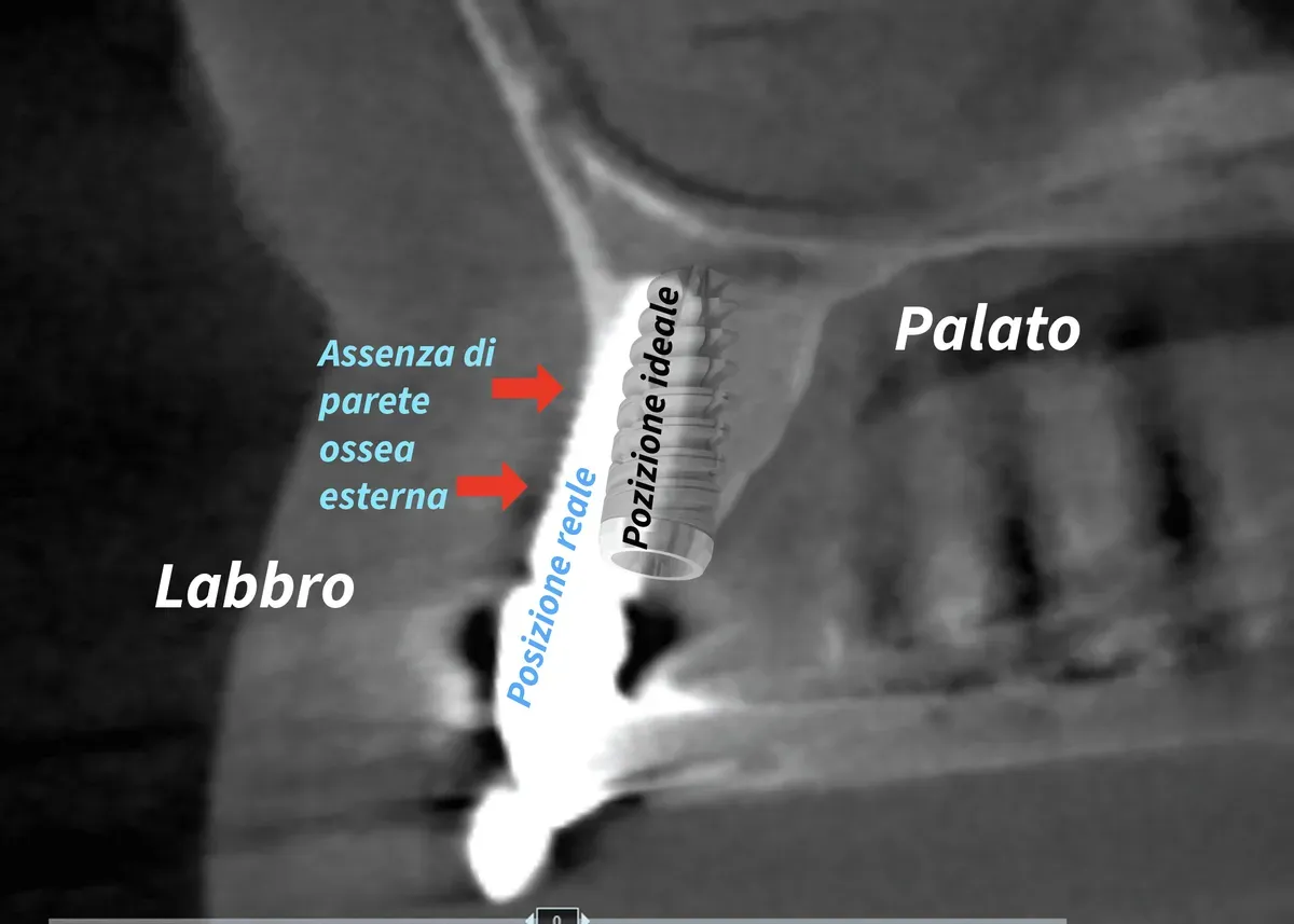
In brief — A malpositioned dental implant is not merely a cosmetic issue, but a biomechanical problem that jeopardizes long-term implant durability. For every 10 degrees of angular deviation, bone resorption risk increases by 0.25 mm. Prevention relies on accurate three-dimensional planning and assessment of the patient’s gingival biotype.
The news comes from SIdP during the 22nd International Congress in Rimini: 2.2 million dental implants were placed in Italy in 2024, but up to 40% turned out to be malpositioned. The numbers are unambiguous. This isn’t a marginal cosmetic issue. Behind the so-called “gummy smile” hides a biomechanical problem that compromises implant longevity and the integrity of peri-implant tissues.
You can read the original article here (Italian): ANSA — boom impianti di denti ma 4 su 10 sono mal messi
Implant malposition isn’t an abstract concept. For every 10 degrees of angular error, bone resorption risk increases by 0.25 millimeters. These numbers tell a precise story of non-axial loading, concentrated stress, and programmed failure of long-term osseointegration.
But what’s the right angle? It depends on the angulation of the teeth and the bone. Because some implants are intentionally placed at an angle — and that’s a perfectly correct, effective long-term technique.
The Biomechanics of Tilted Implants: How Forces Compromise Osseointegration
A dental implant withstands occlusal forces that can reach 60-100 kg during chewing, with peaks above 120 kg on molars. These forces break down into axial and transverse components according to immutable physical principles.
When a single implant is correctly positioned, along the ideal axis of the future crown, the axial component prevails. The load transmits compressively to the surrounding bone through the threads, which function as a load-bearing structure optimized for intrusive forces. The bone responds according to Wolff’s law, remodeling along stress lines and maintaining its density.
A malpositioned dental implant radically changes this biomechanical pattern. Transverse forces increase proportionally with the angle of deviation. It isn’t just geometry — it’s materials mechanics applied to living tissue. Bone tolerates shear forces poorly. Its trabecular structure is built to resist compression along the physiological load lines that formed during the eruption of natural teeth.
Tilted implants, however, work excellently when used correctly with balanced mechanical schemes across multiple units. This is evident in full-arch rehabilitations using the all-on-four or all-on-six technique, where distal tilted implants compensate for a lack of posterior bone and distribute loads through a rigid connecting bar.
Immediate implant placement after extraction is another context where three-dimensional planning matters most, since there’s no residual alveolar wall left to guide positioning.
The problems of implant malposition are always tied to an incorrect, unplanned three-dimensional position — most often translating into a position too far outside the dento-gingival arch. The buccally tilted implant is perhaps the most frequent and devastating complication for peri-implant tissues.
And this happens even with implants that aren’t immediately classifiable as tilted, but which, in practice, point too far outward, compromising the thin buccal bone plate that represents the first protective barrier.
It also happens when a probable resorption isn’t calculated or compensated for during surgery due to concurrent extractions. The loss of bundle bone — the alveolar bone proper that surrounded the natural root — is an inevitable physiological phenomenon that must be anticipated during planning.
Peri-implant Bone Resorption: The Biological Cascade
From an overly outward position to peri-implant bone resorption, the step is short. The process follows a predictable biological sequence that begins with physiological post-surgical bone remodeling.
Gingival biotype plays a critical role in determining the esthetic and functional outcome. According to SIdP data, 60% of the Italian population has constitutionally thin gingiva. In these patients, bone loss quickly translates into gingival recession. Thin buccal bone, typical of the anterior sectors, doesn’t tolerate errors. Once lost, it doesn’t regenerate spontaneously.
The peri-implant biologic width — the complex of soft tissues that seals the interface between implant and oral environment — requires a minimum vertical space of about 3 millimeters. When the implant is too buccal or too shallow, this biologic width cannot establish itself correctly. The result is chronic inflammation of the soft tissues, followed by progressive bone loss.
On the other hand, peri-implant bone resorption can also occur for reasons unrelated to positioning. Peri-implantitis — a bacterial inflammatory disease — is the leading cause of late implant failure. And some systemic therapies radically alter bone metabolism around the implant.
Medications and Implants: What Changes in Bone Remodeling
A malpositioned implant in a healthy patient is a problem. The same implant in a patient taking medications that alter bone metabolism becomes a more urgent problem — because the margin for biological compensation shrinks.
Bisphosphonates and denosumab (Prolia, Xgeva) suppress bone turnover. In a patient with a buccally tilted implant, this means the compensatory remodeling that could stabilize the situation doesn’t happen, or happens insufficiently. The risk of medication-related osteonecrosis of the jaw (MRONJ) is low in osteoporotic patients — only marginally higher than in the general population — but the surgical management of an implant that needs removal changes radically if the patient is on anti-resorptive therapy.
Long-term corticosteroids reduce bone density systemically. An implant with a marginal position that would remain stable for years in a healthy patient can show accelerated bone loss in a patient on chronic corticosteroid therapy.
SSRIs reduce platelet function and — a less well-known fact — some studies suggest a negative effect on bone density through the serotonergic pathway in bone metabolism. Metformin, the world’s most widely prescribed antidiabetic drug, shows a direct effect on osseointegration in animal models, with increased RANKL expression.
There’s one rule: bring the complete list of medications. Not just the “important” ones — all of them. The dentist sees things a general practitioner might not connect.
Three-Dimensional Implant Malposition: Buccal, Palatal and Angular
The term “crooked” is colloquial but clinically ambiguous. An implant can be malpositioned along three spatial dimensions. Each deviation has specific consequences that compromise different aspects of the rehabilitation.
An implant placed too far buccally erodes the thin outer bone plate. In classic implantology (which isn’t gospel but represents consensus built on decades of clinical observation), the minimum safety distance is 1-2 millimeters from the buccal bone profile.
Conversely, an implant placed too far palatally or lingually creates prosthetic and phonetic problems. Crown emergence looks unnatural, home cleaning becomes difficult, esthetics suffer, and speech may be affected by the tongue space taken up. In the upper anterior sectors, a palatal implant forces overcontoured crowns that trap plaque and inflame the tissues.
Angulation is perhaps the most critical parameter in implant malposition. An implant can be angled anteroposteriorly following the natural root axis, but never buccally. Buccal angulation concentrates forces on the outer cortical plate — the thinnest and most vulnerable — accelerating resorption.
I’ve seen too many cases of implants (especially immediately loaded, full-arch cases) with crown emergences pointing outward and total tissue loss in that area. In those cases, they need to be changed. There are no conservative alternatives when the three-dimensional position irreversibly compromises peri-implant biomechanics and biology.
The apico-coronal position, often overlooked, determines whether natural emergence profiles are achievable. An implant placed too shallow exposes the metal threads, while one placed too deep requires extra steps to build crowns with physiological contours (and a conical connection becomes necessary). The general rule places the implant platform 4 millimeters apical to the expected gingival margin, but this is a simplification that must be adapted to the individual biotype and bone profile.
Digital Implant Planning and Computer-Guided Surgery
Modern implant planning starts with a cone-beam CT that provides a volumetric bone map with sub-millimeter resolution. Planning software lets clinicians virtually position the implant considering not just bone anatomy but the final prosthetic project. You can overlay the diagnostic wax-up, see where the crown will emerge, and verify the implant follows the ideal prosthetic axis.
The surgical guide transfers this planning into the operating room. It’s a biocompatible resin template with metal sleeves that guide the drills according to predetermined angulations. Precision runs to tenths of a millimeter for position and degrees for angulation. According to PubMed, a 2025 meta-analysis of 45 studies (Khaohoen et al., Journal of Prosthetic Dentistry) quantifies the advantage: computer-assisted surgery reduces mean angular deviation by 3.87 degrees and apical deviation by 1.10 mm compared with freehand, with robotic surgery achieving the highest clinical accuracy among all protocols tested DOI.
On immediate post-extraction implant placement — where the margin for error is smaller because there’s no alveolar wall to serve as a reference — a 2026 network meta-analysis of 18 studies and 780 implants (Nava et al., Clinical Oral Implants Research) confirms that every guided protocol, static or dynamic, significantly reduces angular deviation compared with freehand, with dynamic and robotic surgery topping the ranking DOI. A second 2026 study on robotic surgery in single-tooth sites (Mahardawi et al., Journal of Dentistry) confirms the robot’s angular advantage but flags that the certainty of evidence remains low to moderate — few studies, a technology still young DOI. Worth knowing before assuming it’s automatically superior.
It isn’t foolproof. Guided surgery requires guide stability during drilling, correct site preparation according to the system’s protocol, and experience with the technique. Errors accumulate: radiographic acquisition error, software segmentation error, guide manufacturing error, intraoperative placement error. Each contributes to the final deviation.
The benefits are many, but the main one is predictability of implant placement. You know in advance where the implant will go. Critical anatomical structures like the mandibular canal or the maxillary sinus can be avoided with calculated safety margins. Flapless surgery — without gingival flap elevation — can also be planned, reducing surgical trauma and speeding healing when the biotype allows it.
Personally, I usually prefer my own method with intraoperative CT, which gets me to the same result with fewer appointments and less stress for the patient. Partly because, in my approach, I usually also regenerate bone and other tissues following the conservative philosophy of bone expansion, the core of the Bonebenders method.
Preoperative biotype assessment should be an integral part of implant planning. Looking at the CT scan isn’t enough. It requires clinical examination of the soft tissues, gingival thickness assessment with transgingival probing, crestal morphology analysis, and periodontal phenotype — which predicts biological response to surgical trauma.
In cases of unfavorable biotype, modifying the biotype itself during surgery should always be considered — through connective-tissue grafts or collagen matrices that increase gingival thickness and protect buccal bone from resorption. When buccal bone shows significant deficits, bone-expansion techniques offer a conservative alternative to traditional guided bone regeneration.
According to PubMed, a 2026 network meta-analysis of 48 RCTs (Cafasso et al., Journal of Periodontal Research) clarifies which technique to choose when soft tissue augmentation is needed around an at-risk implant: an apically positioned flap combined with a free gingival graft remains the most effective option for increasing keratinized mucosa width, while a connective-tissue graft — the bilaminar technique — remains the gold standard for mucosal thickness, with favorable effects on marginal bone level stability DOI. Technique choice matters more than surgical timing.
For Patients: What a Malpositioned Implant Really Means
If you’re considering a dental implant or already have one, it’s important to understand that implant position determines everything else. It isn’t just about where the screw goes into the bone — it’s about where the crown will emerge, how forces will distribute when you chew, and how long your rehabilitation will last.
A well-positioned implant blends invisibly into the arch, cleans easily, doesn’t trap plaque, and keeps the surrounding gingiva healthy and pink.
A malpositioned implant, on the other hand, can trigger a cascade of problems: gingiva retracting to expose the metal, a crown that protrudes or sits too far back, difficulty flossing, chronic inflammation, and the need for costly corrective procedures.
Prevention comes down to choosing a professional who uses modern planning technology, dedicates time to the diagnostic phase, and shows you on screen where the implant will go before starting.
The apparently higher upfront cost of accurate planning pays for itself over time through fewer complications and longer implant survival.
What to Actually Do About a Malpositioned Dental Implant
The first thing to do if you suspect a malpositioned implant is a specialist clinical and radiographic evaluation. Not every situation requires removal. The path depends on three variables: extent of the deviation, condition of the peri-implant tissues, and prosthetic correctability.
| Situation | Clinical option | Timing |
|---|---|---|
| Mild angular deviation (<15°), healthy tissues (often intentional for anatomical reasons, not an error) | Prosthetic compensation with an angled abutment | Immediate, no surgery |
| Buccal (outward) position with thin gingiva | Connective-tissue graft to thicken the biotype | Before recession appears |
| Buccal (outward) position with early bone loss | Guided bone regeneration + possible prosthetic correction | Within 6-12 months of placement |
| Severely incorrect position with advanced bone loss | Implant removal, regeneration, reinsertion in the correct position | Careful planning, 6-18 months total |
The practical rule: the earlier you intervene, the more conservative options remain available. A buccally tilted implant caught at 3 months, before the final restoration, is manageable with minimal intervention. The same implant with 3 years of progressive bone loss requires a far more complex treatment plan.
Repositioning a Dental Implant: When It’s Necessary and How It’s Done
Implant repositioning isn’t a single procedure but a pathway with three distinct phases. The first is removal of the malpositioned implant: performed with dedicated instruments (trephine or reverse-torque kit), under local anesthesia, preserving as much surrounding bone as possible. A well-osseointegrated implant requires more force to remove than one with peri-implant bone loss.
The second phase is regeneration of the site. The socket left by the removed implant and any bone defect are treated with autologous bone, xenografts, or synthetic bone substitutes, protected by membranes. Healing times range from 4 to 9 months depending on the extent of the defect and the technique used. In this context, the conservative bone-expansion approach can significantly shorten timelines compared with classic guided bone regeneration.
The third phase is reinsertion in the correct position of a new implant, preceded by fresh three-dimensional planning that accounts for the regenerated bone morphology. In selected cases, when the site is favorable and bone quality is adequate, a single-session approach is possible: removal, regeneration, and immediate reimplantation. This option requires experience and rigorous case selection.
The cost of implant repositioning varies and depends on the complexity of the bone defect, the need for regeneration, and the materials used. In general terms, the full pathway costs about the same as a new implant with bone regeneration — a significant expense that accurate initial planning could have avoided.
Conclusions: Precision as an Ethical Imperative
The SIdP data paint a concerning but unsurprising picture. Implantology has become a widespread, accessible, almost routine practice. The technique is well established, the materials are reliable, and osseointegration success rates are high. But these results come from studies conducted in referral centres, by experienced operators, on patients selected under rigorous inclusion criteria.
In daily practice, reality is more complex. Difficult anatomies, patients with comorbidities and chronic therapies, high esthetic expectations, limited budgets. The temptation to simplify is strong. Placing the implant where it’s easier rather than where it’s right. Accepting a compromise in implant position to avoid a bone regeneration procedure that lengthens treatment time and raises costs.
These compromises are paid for over time with predictable complications: peri-implant bone resorption, gingival recession, peri-implantitis, esthetic and functional failures.
The technology available today makes it possible to minimize implant malposition errors. Cone-beam CT provides a precise surgical map of three-dimensional anatomy. Planning software allows accurate simulations of the final result. For those who favor them, computer-guided surgical guides are an excellent aid for transferring virtual planning into clinical reality.
The message to patients is clear: a malpositioned dental implant isn’t just a cosmetic issue. It’s a biomechanical problem that compromises the longevity of the rehabilitation. Prevention comes through accurate planning, appropriate technology, and clinical experience in managing bone deficits and soft peri-implant tissues.
The higher upfront cost of correct implant planning pays for itself over time through fewer complications and longer implant survival. The quality of the positioning determines the quality of life with the implant for decades to come.
For more: visit dentipiu.it for clinical information, or read the scientific literature on implant malposition and peri-implant bone loss.
References
- Khaohoen A, Powcharoen W, Yoda N, Rungsiyakull C, Rungsiyakull P. Accuracy in dental implant placement: a systematic review and meta-analysis comparing computer-assisted (static, dynamic, robotics) and noncomputer-assisted (freehand, conventional guide) approaches. J Prosthet Dent. 2025;134(1):91.e1-91.e25. doi:10.1016/j.prosdent.2025.03.038 · PMID: 40221370
- Nava P, Sabri H, Hazrati P, Nava C, Saleh MHA, Wang HL. Accuracy of static, dynamic, and robotic guided surgery in immediate implant placement: a systematic review and network meta-analysis. Clin Oral Implants Res. 2026;37(5):525-542. doi:10.1111/clr.70100 · PMID: 41714848
- Mahardawi B, Jiaranuchart S, Tajmiri G, Mattheos N, Pimkhaokham A. Does robotic surgery offer the highest accuracy for delayed implant placement in single-tooth spaces? A network meta-analysis of randomized clinical trials. J Dent. 2026;106876. doi:10.1016/j.jdent.2026.106876 · PMID: 42385837
- Cafasso E, Baima G, Campagna A, Tavelli L, Aimetti M. Soft tissue augmentation around dental implants: techniques, timing, and comparative efficacy — a systematic review and network meta-analysis. J Periodontal Res. 2026;61(4):332-354. doi:10.1111/jre.70100 · PMID: 41858220
FAQ
How long does a malpositioned dental implant last?
Can a crooked implant be corrected?
What are the symptoms of a malpositioned implant?
Does a malpositioned implant cause pain?
How do you know if an implant is malpositioned?
Does a malpositioned dental implant always need to be removed?
How much does it cost to remove and reposition a dental implant?
The implant moves slightly: is it malpositioned?
Can medications affect an implant already in place?
Cite this article
Ready for theses, slides and papers. If you cite it on a web page, make the URL a link so readers can reach the source.
Bruschi E. Malpositioned Dental Implant: Risks, Symptoms and Solutions [Internet]. Frosinone (Italy): Bonebenders; 2025 [updated 2026 Jul 9; cited 2026 Oct 25]. Available from: https://bonebenders.com/en/blog/malpositioned-dental-implant-biomechanical-risks/ Bruschi, E. (2025, November 29). Malpositioned Dental Implant: Risks, Symptoms and Solutions. Bonebenders. https://bonebenders.com/en/blog/malpositioned-dental-implant-biomechanical-risks/
@misc{bruschi2025malpositioned,
  author       = {Bruschi, Ernesto},
  title        = {Malpositioned Dental Implant: Risks, Symptoms and Solutions},
  year         = {2025},
  month        = nov,
  howpublished = {Bonebenders},
  url          = {https://bonebenders.com/en/blog/malpositioned-dental-implant-biomechanical-risks/},
  note         = {Accessed 2026-10-25}
} Go deeper
Espansione crestale (split crest) →Allargare la cresta senza trapianto — tecnica ERE e bonebending
Need a professional opinion?
Book an appointment at Dr. Bruschi's practice in Frosinone. First visit includes full diagnosis and personalised treatment plan.
Stai valutando un impianto dentale?
Ti hanno detto che non hai abbastanza osso, o ti hanno proposto un’arcata fissa? Ho scritto una guida di dieci pagine: le strade possibili, gli esami che servono e le dieci domande da fare prima di firmare.
Ricevi la guidaStay Updated
New articles on periodontology, implantology and oral surgery — delivered to your inbox.
Comments
Loading comments...
Leave a comment