Proteins That Trigger Osseointegration: 3 Phases
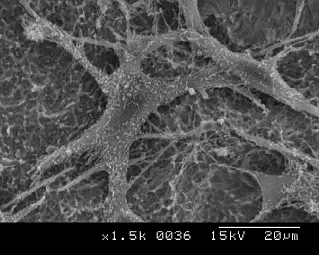
In brief — Osseointegration starts with blood: within seconds proteins coat titanium, then cells arrive, then bone grows. Three phases. Surface matters — yet recent clinical data shrink the promise of “super-hydrophilic” marketing.
The moment the implant meets blood, the titanium surface stops being only metal.
Within seconds albumin, fibrinogen and immunoglobulins wet it. That light protein film decides who comes next: bone-building cells, macrophages that steer healing, or fibroblasts — and there you often end in fibrointegration: soft tissue around the implant, not bone.
Material still matters — purity, sterility, shape. But if you stop at the commercial brand promising miracles of purity, you miss the point. Osseointegration is born mainly from what happens on the surface, in the interaction between the patient’s blood and titanium.
An impure surface, of course, does not help.
Phase 1: blood arrives first
Blood wets the implant. Plasma proteins bind. All in a handful of seconds.
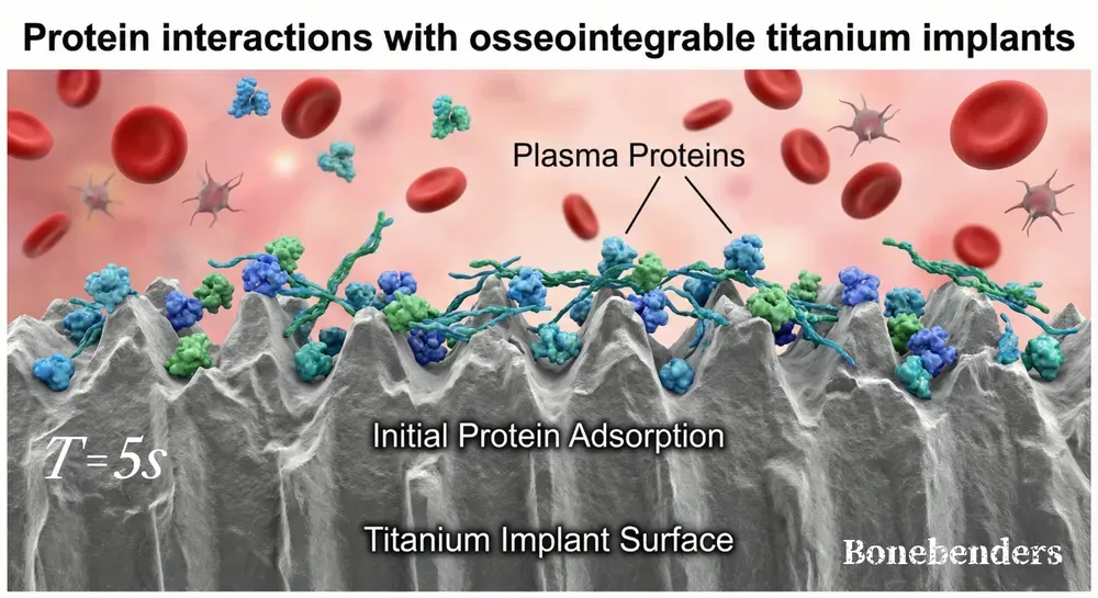
More wettable surfaces help: fewer air bubbles, a more continuous protein film. Liu and colleagues (BMC Oral Health, 2025) saw that a TiO₂ nanowire treatment makes titanium wetter and holds proteins better; in the lab, bone cells respond more readily. I covered the same window in “Bind me!” Blood and Titanium: the first minutes set what follows.
A practical detail to watch. Çiftçi and colleagues (BMC Oral Health, 2026; 54 studies) reviewed what happens if saliva touches the implant during surgery: the surface wets worse, bone cells attach less, and no rinse brings titanium back to its starting state. Better not to contaminate.
That first protein layer is the messenger. Cells read the molecules spread over the metal.
Phase 2: cells recognize and attach
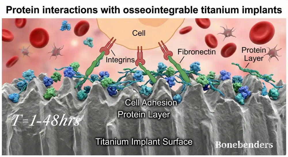
Phase-1 proteins hook integrins — small “latches” on the membrane of cells that approach. Fibronectin bridges the gap. Without that bridge, cells struggle to stay.
Komatsu, Matsuura, Suzumura and Ogawa (Materials Today Bio, 2023) placed osteoblasts on smooth and microrough titanium and read which genes switch on. On the rough surface, immune-related genes rose more, and with them the production of proteins that help cell attachment — laminin, fibronectin, thrombospondin — and those that organize the cell’s cytoskeleton (helping movement toward the new surface). That is one reason a macro- and micro-structured surface favors osseointegration.
In parallel, macrophages work. At first they push inflammation, as in a foreign-body reaction; then, if things go well, they shift to a more “repair” profile and favor new bone and vessels. Stem cells and osteoblast precursors arrive too. If myofibroblasts win, you get fibrointegration: connective tissue instead of bone.
Mesa-Restrepo and colleagues (ACS Biomaterials Science & Engineering, 2024) traced the full chain: proteins that bind → the body’s response to a foreign object → how the immune system steers healing. Chemistry, electric charge, wettability and roughness decide which proteins stay and how they fold. Cells “see” that shape. In clinical trials this variable is still barely measured.
This phase runs from the first hours through 24–48 hours.
Phase 3: contact with bone begins
Once precursors of osteoblasts have attached, they grow into mature cells. Bone does not touch “bare” titanium: a protein anchorage remains, on which osteoblasts lay matrix, then calcium crystals (hydroxyapatite).
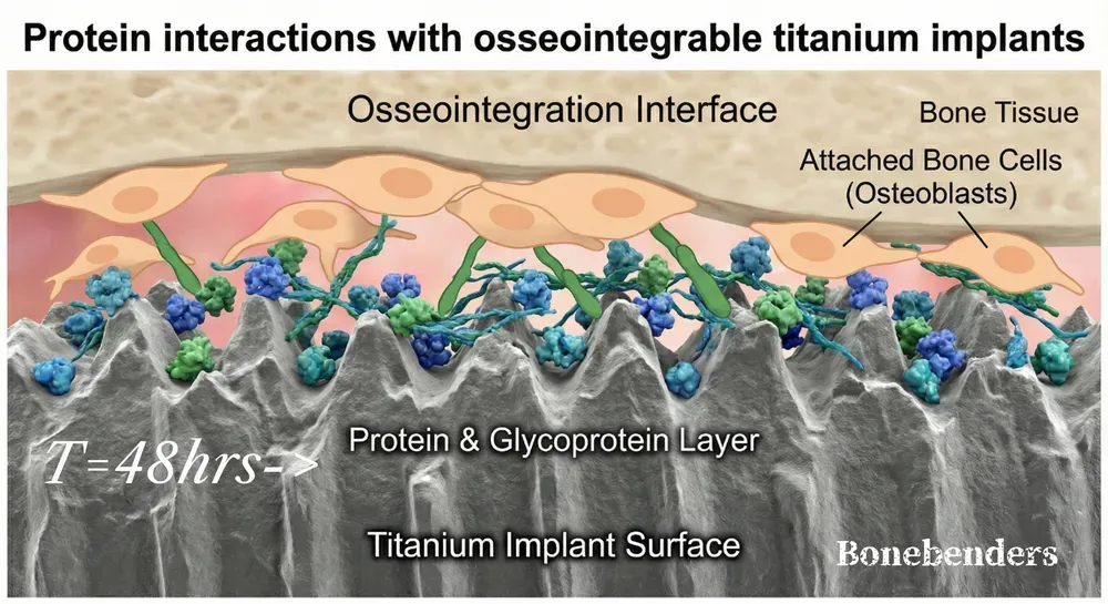
Days, then weeks. From there on, bone around the implant keeps renewing — the same living network described in the osteocyte connectome.
Ivanovski, Lee, Fernandez-Medina, Pinto, Andrade and Quirynen (Periodontology 2000, 2025) studied the effects of the patient’s platelet concentrates (PRP, L-PRF and similar): the idea is to put host proteins on titanium so new bone starts sooner. The results, at least in that paper, are not striking. PRP and liquid fibrinogen help little in clinic. L-PRF membranes in the osteotomy site may give more stability and less marginal bone loss in the first six weeks. After that, outcomes look the same.
Surface and clinical success
Outcome depends on the start. Rough, hydrophilic surfaces are meant to hold blood and the protein film faster.
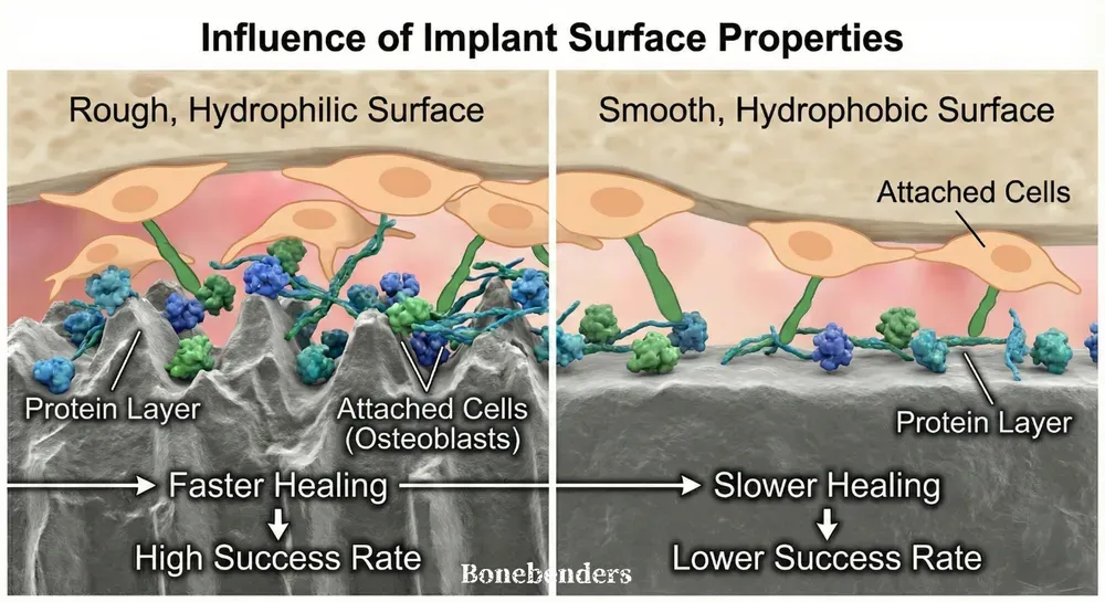
In clinic the picture is tighter than the brochure. Canullo, Menini, Guardone, Merlini, Cameroni, Sculean, Pesce and Del Fabbro (Oral Health & Preventive Dentistry, 2025) pooled 13 randomized and 2 non-randomized studies — 1,256 implants in 596 patients — comparing “bioactive” or super-hydrophilic surfaces with traditional ones. At one year, survival and marginal bone loss: the same. Measured stability (ISQ) was similar at placement and at one month; at three months bioactive surfaces gained a little more. The authors say it plainly: a “bioactive” surface alone does not justify immediate or early loading.
Vílchez, Caneiro, Lima, Sanz-Sánchez, Montero, Figuero, Blanco and Sanz (Clinical Oral Implants Research, 2026) ran a multicenter split-mouth study: 68 patients, 136 implants, modified hydrophilic SLA versus conventional SLA. At 12 months after loading, marginal bone loss was practically the same (0.04 mm vs 0.07 mm). Soft tissue, stability, healing: overlapping. Two different surfaces. Same one-year outcome.
I have already written why smooth surfaces and implant shape alone are not enough: placement stability, tissue handling and patient metabolism still decide. Proteins open the door. Who walks through — and who stays — depends on the concrete case.
Three to six months for solid integration remains the rule. Speeding the first two phases makes sense. Promising more from the surface alone is a leap recent data do not support.
Osseointegration is a dialogue among blood, proteins and surface. Titanium is the support. Biology writes the rest.
FAQ
What are the three biological phases of titanium implant osseointegration?
Why does titanium implant surface matter for osseointegration?
What happens in the first phase, and why does it matter?
How do cells recognize and attach to the implant surface?
How long does osseointegration take to complete?
References
- Ivanovski S, Lee RSB, Fernandez-Medina T, et al. Impact of autologous platelet concentrates on the osseointegration of dental implants. Periodontol 2000. 2025;97(1):271-286. doi:10.1111/prd.12563 · PMID: 38647020 · free full text
- Canullo L, Menini M, Guardone L, et al. Do Super-hydrophilic Surfaces Affect Implant Primary Stability in the Early Healing Phase of Osseointegration? A Systematic Review with Metanalysis. Oral Health Prev Dent. 2025;23:469-478. doi:10.3290/j.ohpd.c_2235 · PMID: 40853239 · free full text
- Vílchez B, Caneiro L, Lima C, et al. Clinical and Radiographic Performance of Two Distinct Sandblasted, Large-Grit, Acid-Etched Implant Surfaces: A Split-Mouth Randomized Clinical Trial. Clin Oral Implants Res. 2026;37(2):141-154. doi:10.1111/clr.70071 · PMID: 41199413 · free full text
- Mesa-Restrepo A, Byers E, Brown JL, et al. Osteointegration of Ti Bone Implants: A Study on How Surface Parameters Control the Foreign Body Response. ACS Biomater Sci Eng. 2024;10(8):4662-4681. doi:10.1021/acsbiomaterials.4c00114 · PMID: 39078702
- Komatsu K, Matsuura T, Suzumura T, et al. Genome-wide transcriptional responses of osteoblasts to different titanium surface topographies. Mater Today Bio. 2023;23:100852. doi:10.1016/j.mtbio.2023.100852 · PMID: 38024842 · free full text
- Liu Y, Yang J, Guo Y, et al. Photocatalytic cleaning effect of TiO<sub>2</sub> nanowires increase the osteoconductivity of the titanium surface. BMC Oral Health. 2025;25(1):1886. doi:10.1186/s12903-025-07294-3 · PMID: 41299395 · free full text
- Çi̇ftçi̇ S, Kocamemi̇k S, Aktaş KA, et al. Salivary contamination during implant placement: effects on titanium surface properties and early osseointegration- a systematic review. BMC Oral Health. 2026;26(1):1015. doi:10.1186/s12903-026-08289-4 · PMID: 42015160 · free full text
- Ivanovski S, Lee RSB, Fernandez-Medina T, et al. Impact of autologous platelet concentrates on the osseointegration of dental implants. Periodontol 2000. 2025;97(1):271-286. doi:10.1111/prd.12563 · PMID: 38647020 · free full text
- Canullo L, Menini M, Guardone L, et al. Do Super-hydrophilic Surfaces Affect Implant Primary Stability in the Early Healing Phase of Osseointegration? A Systematic Review with Metanalysis. Oral Health Prev Dent. 2025;23:469-478. doi:10.3290/j.ohpd.c_2235 · PMID: 40853239 · free full text
- Vílchez B, Caneiro L, Lima C, et al. Clinical and Radiographic Performance of Two Distinct Sandblasted, Large-Grit, Acid-Etched Implant Surfaces: A Split-Mouth Randomized Clinical Trial. Clin Oral Implants Res. 2026;37(2):141-154. doi:10.1111/clr.70071 · PMID: 41199413 · free full text
- Mesa-Restrepo A, Byers E, Brown JL, et al. Osteointegration of Ti Bone Implants: A Study on How Surface Parameters Control the Foreign Body Response. ACS Biomater Sci Eng. 2024;10(8):4662-4681. doi:10.1021/acsbiomaterials.4c00114 · PMID: 39078702
- Komatsu K, Matsuura T, Suzumura T, et al. Genome-wide transcriptional responses of osteoblasts to different titanium surface topographies. Mater Today Bio. 2023;23:100852. doi:10.1016/j.mtbio.2023.100852 · PMID: 38024842 · free full text
Cite this article
Ready for theses, slides and papers. If you cite it on a web page, make the URL a link so readers can reach the source.
Bruschi E. Proteins That Trigger Osseointegration: 3 Phases [Internet]. Frosinone (Italy): Bonebenders; 2026 [updated 2026 Aug 6; cited 2026 Oct 2]. Available from: https://bonebenders.com/en/blog/proteins-that-trigger-osseointegration-3-phases/ Bruschi, E. (2026, January 6). Proteins That Trigger Osseointegration: 3 Phases. Bonebenders. https://bonebenders.com/en/blog/proteins-that-trigger-osseointegration-3-phases/
@misc{bruschi2026proteins,
  author       = {Bruschi, Ernesto},
  title        = {Proteins That Trigger Osseointegration: 3 Phases},
  year         = {2026},
  month        = jan,
  howpublished = {Bonebenders},
  url          = {https://bonebenders.com/en/blog/proteins-that-trigger-osseointegration-3-phases/},
  note         = {Accessed 2026-10-02}
} Go deeper
Espansione crestale (split crest) →Allargare la cresta senza trapianto — tecnica ERE e bonebending
Need a professional opinion?
Book an appointment at Dr. Bruschi's practice in Frosinone. First visit includes full diagnosis and personalised treatment plan.
Stai valutando un impianto dentale?
Ti hanno detto che non hai abbastanza osso, o ti hanno proposto un’arcata fissa? Ho scritto una guida di dieci pagine: le strade possibili, gli esami che servono e le dieci domande da fare prima di firmare.
Ricevi la guidaStay Updated
New articles on periodontology, implantology and oral surgery — delivered to your inbox.
Comments
Loading comments...
Leave a comment