Myofibroblasts Close the Wound, Then They Have to Leave
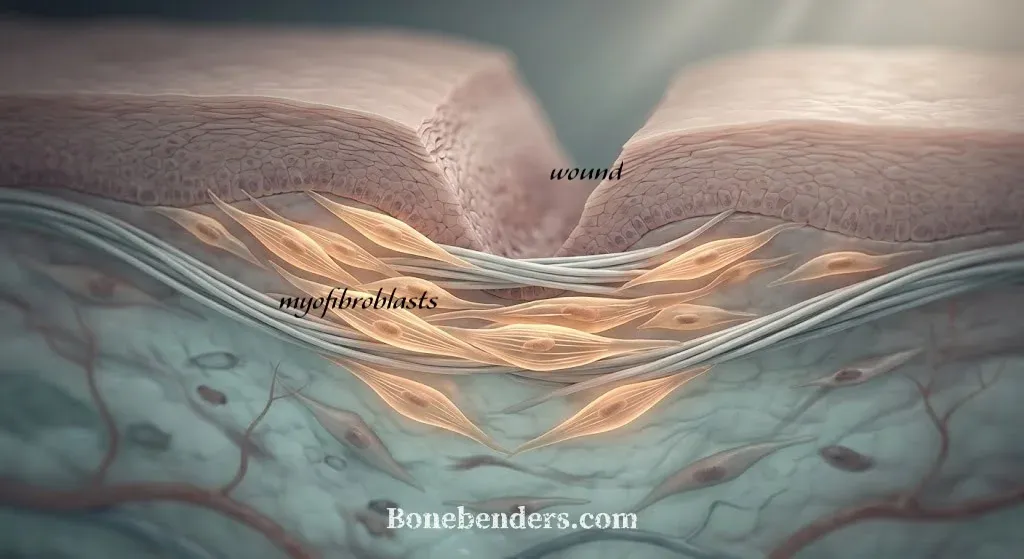
In brief — Myofibroblasts are activated fibroblasts that close wounds, oral wounds included, by pulling the edges together and laying down collagen. They do the same job around an implant, and their activity is necessary. Healing succeeds only when, once the work is done, they die by apoptosis or return to quiescence. When they stay active, the tissue becomes fibrotic. Mechanical tension is the signal that keeps them alive, and the surgeon controls it with his own hands.
Seven Days of Cellular Tension
Remove the sutures at seven days and the mucosa is closed. Little or no visible scar. Without myofibroblasts, closure would be far less effective.
Most of that week’s work was done by cells that, three days earlier, were quiet fibroblasts asleep in the connective tissue. The surgical wound woke them up. They built a contractile apparatus inside themselves, pulled the wound edges toward each other, and filled the gap by producing and depositing new collagen. Then they vanished.
Their name is myofibroblasts. I’ve already written about their dark side, fibrous encapsulation. Here I want to talk about the useful side, the one we rely on every day without naming it, and about the final step that decides everything: when the job is done and they exit the stage.
What a Fibroblast Becomes When You Wound It
Younesi, Miller, Barker, Rossi, and Hinz published the review that now serves as the reference point for this cell, in 2024 in Nature Reviews Molecular Cell Biology. The path they describe has two stages.
First stage. The fibroblast senses that the matrix around it has changed consistency. How does it sense this? Probably through a mix of biochemical and mechanical communication, proximity and transmission, the kind of thing I keep coming back to on this blog.
The wound disrupts the tissue that had kept the cell relaxed and dormant, replacing it with a fibrin clot and then a repair-stage granulation tissue. The cell responds by forming stress fibers and new focal adhesions in the healing extracellular matrix. Hinz calls this stage the proto-myofibroblast. And the proto-myofibroblast pulls, but only a little.
Second stage. A second signal is needed: TGF-β1 (transforming growth factor beta 1), a multifunctional cytokine that regulates growth, proliferation, differentiation, and cell death.
Here’s the remarkable part. TGF-β1 is already there, in the matrix, kept latent, bound to a protein that holds it inactive. The fibroblast releases it with its own membrane integrins, by pulling. The stiffer the matrix, the harder the cell pulls, the more TGF-β is released, the more active the cell becomes. It’s a self-feeding loop. At that point the cell expresses smooth-muscle actin, α-SMA, and becomes a full myofibroblast. The force it generates rises sharply.
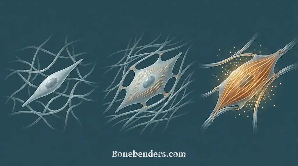
You could say that quiescent fibroblasts and latent TGF-β1 form a first line of defense for connective tissue, ready to activate like a fast emergency service the moment a trauma or wound of any kind occurs.
Schuster, Younesi, Ezzo, and Hinz, in Cold Spring Harbor Perspectives in Biology in 2023, use a definition I like. They call myofibroblasts the construction workers of repair. They produce the matrix, organize it, put it under tension to close the wound. A construction site with no workers never closes.
A construction site where the workers never leave once the job is finished becomes a different problem. I’ll get to that.
Oral Mucosa Heals Better Than Skin, Myofibroblasts Included
Iglesias-Bartolome and colleagues, in Science Translational Medicine in 2018, did something simple and rare. They made identical wounds in the mouth and on the arm of the same volunteers, and took sequential biopsies. Oral wounds closed much faster. The oral mucosa already had the genetic repair programs switched on at rest. Factors like SOX2 and PITX1 keep the epithelium ready to begin repair before the surgeon even arrives.
The data on myofibroblasts comes from an animal model that closely resembles humans. Mak, Larjava, Häkkinen, and colleagues, in 2009 in the Journal of Dermatological Science, compared mucosal and skin wounds in the red Duroc pig, an animal that scars hypertrophically the way we do. In the mouth, inflammation resolved sooner, myofibroblasts appeared and disappeared sooner, and the scar stayed minimal. In skin, they stayed active much longer, and the scar grew much larger.
One molecular detail sharpens the difference further. Nikoloudaki, Creber, and Hamilton, in 2020 in the American Journal of Physiology-Cell Physiology, studied periostin, a matrix protein. In skin, periostin drives fibroblasts to become myofibroblasts, and this molecule is abundant in scars.
In gum tissue, periostin regulates matrix synthesis in a similar way but doesn’t push cells into persistent myofibroblast activity. Same protein, two tissues, two slightly different fates.
Vescarelli, Pilloni, and colleagues, in 2017 in the Journal of Clinical Periodontology, compared two tissues from the same mouth: oral mucosa and attached gingiva. Only in the mucosa does healing switch on autophagy, the process cells use to recycle their own internal components, and only there do smooth-muscle actin and type I collagen rise. In attached gingiva, autophagy stays off, myofibroblasts differentiate less, and no scar forms. Autophagy turns out to be one of the levers that decides, inside the same mouth, whether a tissue scars.
What does this biological mechanism mean for those of us who operate?
It tells us, unmistakably, that oral mucosa already has an internal system to activate myofibroblasts early and switch them off early. The surgeon can work with that system, to his own advantage and his patients’.
For those of us in the Bonebenders world, myofibroblasts and their peculiar behavior in oral mucosal healing are the foundation of second-intention healing, a technique we hold dear. Without these remarkable cells, the wide osteo-mucosal regenerations that set our work apart wouldn’t be possible.
After an Extraction, and Around an Implant
An empty socket is a deep wound with bony walls. The clot is replaced by repair-stage granulation tissue, and while osteoblasts start laying down bone at the bottom, myofibroblasts at the surface pull the gum margins toward the center. Without them, the socket would stay open far longer.
Around an implant the same thing happens, with one difference. Sculean, Gruber, and Bosshardt, in their 2014 review in the Journal of Clinical Periodontology, describe the peri-implant mucosa that forms after healing: a connective tissue poor in cells and vessels, rich in collagen, with fibers running parallel to the titanium surface. It’s a scar-like tissue, histologically speaking. Around a natural tooth, by contrast, the fibers insert into cementum and anchor directly. The implant has no layer of “root cementum” cells, and the tissue can only appose itself as closely as possible.
Peri-implant connective tissue is the product of myofibroblasts. The peri-implant biological seal that results works, as long as nothing disturbs it in the first weeks of formation. Every time the flap is lifted, every time a healing screw is swapped, the construction site reopens and the workers come back.
The bone side of the story is more delicate. In a socket, excessive early contraction of the granulation tissue may contribute to ridge resorption. It’s one of several hypotheses that have circulated for years about post-extraction resorption.
The Hardest Part for a Myofibroblast: Leaving
Desmoulière, Redard, Darby, and Gabbiani, in 1995 in the American Journal of Pathology, showed that the transition from repair-stage granulation tissue to scar happens by apoptosis, a genetically programmed and controlled form of cell death. Cells disappear in waves from the repair site, and myofibroblasts go first. Thirty years later, the Younesi and Hinz review adds two alternatives to programmed death: some myofibroblasts revert to ordinary fibroblasts, others enter a state of unproductive senescence.
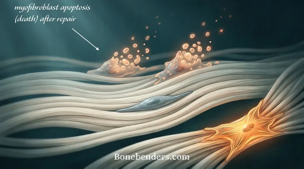
What decides their fate is almost always the same signal that switched them on. If tension in the granulation tissue drops, the TGF-β loop breaks and the cell loses its reason to exist. If tension stays, the cell stays. And here’s a concept I find fascinating: mechanical memory. A fibroblast kept on a stiff matrix for a long time retains epigenetic modifications that keep it primed to reactivate, even after the stiffness is gone.
An implant is undeniably a rigid matrix by definition. Noskovicova, Hinz, and Pakshir wrote as much in Cells in 2021. The rigidity of implantable devices is itself a stimulus that keeps myofibroblasts active, and the fibrous capsule that wraps pacemakers and breast implants is the result of this biological response. In our field, osteoblasts reach the surface quickly, before fibrosis can organize, and they win the race. That’s osseointegration. But the race is real, and it’s why the first week on titanium matters as much as, or more than, the months that follow.
What Changes in Practice
Gao and colleagues, in 2026 in The American Surgeon, reviewed the literature on wound contraction with a clear goal: giving the surgeon a practical guide. Their conclusion is unambiguous. Contraction of moderate intensity and duration helps healing. Excessive or prolonged contraction ruins it. Intensity and duration depend on how many myofibroblasts are present and how long they stay active.
Here are the practical implications for oral surgery:
- Passive closure of mucosal flaps. A tightly sutured flap is a tissue under tension, and tension is the signal that keeps myofibroblasts active past the point they’re needed. Sutures should bring edges together gently, not pull them.
- Primary implant stability. An implant that moves even slightly, micromotion, transmits a cyclic load to the clot. Micromotion is the mechanical signal that favors the fibroblastic line over the osteoblastic one. This data is old, and it’s solid.
- Periosteum. It’s the site’s cell reserve and the tissue best able to bear tension. Fully detaching it, or leaving it torn, shifts the work onto the mucosa’s myofibroblasts, which pull but don’t build bone.
- Don’t reopen implants or unscrew abutments more than necessary. Every reopening restarts the activation cycle. Fewer reopenings, fewer cycles, a more stable tissue. This also applies to component changes in the first weeks.
- Patients who switch off myofibroblasts poorly. Uncontrolled diabetes, smoking, chronic corticosteroids: these alter inflammation, and with it the phase in which myofibroblasts should retreat from the surgical site. In these patients, the rules above matter twice as much.
None of these rules is new. Myofibroblast biology explains why they work, and gives you a way to judge when a shortcut will cost you.
Three Paths Forward
You can keep suturing the way you always have. Most of the time, oral mucosa, with its perpetually active healing program, forgives you.
You can go back over your technique looking for the points where you introduce tension without noticing. It happens with short flaps, release incisions kept too conservative, forced closures over grafts that are too large, tunnel techniques being a common case. This is the path I recommend most.
Or you can read the Hinz review, which is long and not easy, but which can help you look at your next procedure with different eyes.
Remember that once seven days have passed, it’s best for the workers to go home.
References
- Younesi FS, Miller AE, Barker TH, Rossi FMV, Hinz B. Fibroblast and myofibroblast activation in normal tissue repair and fibrosis. Nat Rev Mol Cell Biol. 2024;25(8):617-638. doi:10.1038/s41580-024-00716-0 · PMID: 38589640
- Schuster R, Younesi F, Ezzo M, Hinz B. The role of myofibroblasts in physiological and pathological tissue repair. Cold Spring Harb Perspect Biol. 2023;15(1):a041231. doi:10.1101/cshperspect.a041231 · PMID: 36123034
- Iglesias-Bartolome R, Uchiyama A, Molinolo AA, Abusleme L, Brooks SR, Callejas-Valera JL, et al. Transcriptional signature primes human oral mucosa for rapid wound healing. Sci Transl Med. 2018;10(451):eaap8798. doi:10.1126/scitranslmed.aap8798 · PMID: 30045979
- Mak K, Manji A, Gallant-Behm C, Wiebe C, Hart DA, Larjava H, et al. Scarless healing of oral mucosa is characterized by faster resolution of inflammation and control of myofibroblast action compared to skin wounds in the red Duroc pig model. J Dermatol Sci. 2009;56(3):168-180. doi:10.1016/j.jdermsci.2009.09.005 · PMID: 19854029
- Nikoloudaki G, Creber K, Hamilton DW. Wound healing and fibrosis: a contrasting role for periostin in skin and the oral mucosa. Am J Physiol Cell Physiol. 2020;318(6):C1065-C1077. doi:10.1152/ajpcell.00035.2020 · PMID: 32267719
- Vescarelli E, Pilloni A, Dominici F, Pontecorvi P, Angeloni A, Polimeni A, Ceccarelli S, Marchese C. Autophagy activation is required for myofibroblast differentiation during healing of oral mucosa. J Clin Periodontol. 2017;44(10):1039-1050. doi:10.1111/jcpe.12767 · PMID: 28646601
- Sculean A, Gruber R, Bosshardt DD. Soft tissue wound healing around teeth and dental implants. J Clin Periodontol. 2014;41 Suppl 15:S6-S22. doi:10.1111/jcpe.12206 · PMID: 24641001
- Desmoulière A, Redard M, Darby I, Gabbiani G. Apoptosis mediates the decrease in cellularity during the transition between granulation tissue and scar. Am J Pathol. 1995;146(1):56-66. PMID: 7856739
- Gao S, Tu L, Feng Z, Chen H, Lin J, Gao W. Advances in the mechanism of myofibroblasts in wound contraction: clinical implications for surgical practice. Am Surg. 2026;92(8):2212-2218. doi:10.1177/00031348261423914 · PMID: 41655060
- Noskovicova N, Hinz B, Pakshir P. Implant fibrosis and the underappreciated role of myofibroblasts in the foreign body reaction. Cells. 2021;10(7):1794. doi:10.3390/cells10071794 · PMID: 34359963
FAQ
What is a myofibroblast, in one sentence?
Do myofibroblasts also work around a dental implant?
If the mouth heals without scarring, why is the gum tissue around an implant a scar-like tissue?
Can a myofibroblast that pulls too hard cause bone loss after an extraction?
Isn't this all lab theory? In practice, all that matters is a stable implant.
Are there drugs that switch off myofibroblasts?
References
- https://doi.org/10.1038/s41580-024-00716-0
- https://pubmed.ncbi.nlm.nih.gov/38589640/
- https://doi.org/10.1101/cshperspect.a041231
- https://pubmed.ncbi.nlm.nih.gov/36123034/
- https://doi.org/10.1126/scitranslmed.aap8798
- https://pubmed.ncbi.nlm.nih.gov/30045979/
- https://doi.org/10.1016/j.jdermsci.2009.09.005
- https://pubmed.ncbi.nlm.nih.gov/19854029/
- https://doi.org/10.1152/ajpcell.00035.2020
- https://pubmed.ncbi.nlm.nih.gov/32267719/
- https://doi.org/10.1111/jcpe.12767
- https://pubmed.ncbi.nlm.nih.gov/28646601/
- https://doi.org/10.1111/jcpe.12206
- https://pubmed.ncbi.nlm.nih.gov/24641001/
- https://pubmed.ncbi.nlm.nih.gov/7856739/
- https://doi.org/10.1177/00031348261423914
- https://pubmed.ncbi.nlm.nih.gov/41655060/
- https://doi.org/10.3390/cells10071794
- https://pubmed.ncbi.nlm.nih.gov/34359963/
Cite this article
Ready for theses, slides and papers. If you cite it on a web page, make the URL a link so readers can reach the source.
Bruschi E. Myofibroblasts Close the Wound, Then They Have to Leave [Internet]. Frosinone (Italy): Bonebenders; 2026 [cited 2026 Sep 21]. Available from: https://bonebenders.com/en/blog/myofibroblasts-oral-surgery-implant-healing/ Bruschi, E. (2026, September 20). Myofibroblasts Close the Wound, Then They Have to Leave. Bonebenders. https://bonebenders.com/en/blog/myofibroblasts-oral-surgery-implant-healing/
@misc{bruschi2026myofibroblasts,
  author       = {Bruschi, Ernesto},
  title        = {Myofibroblasts Close the Wound, Then They Have to Leave},
  year         = {2026},
  month        = sep,
  howpublished = {Bonebenders},
  url          = {https://bonebenders.com/en/blog/myofibroblasts-oral-surgery-implant-healing/},
  note         = {Accessed 2026-09-21}
} Go deeper
Espansione crestale (split crest) →Allargare la cresta senza trapianto — tecnica ERE e bonebending
Need a professional opinion?
Book an appointment at Dr. Bruschi's practice in Frosinone. First visit includes full diagnosis and personalised treatment plan.
Stai valutando un impianto dentale?
Ho scritto una guida in 8 capitoli che spiega tutto quello che un paziente dovrebbe sapere prima di sedersi in poltrona. Niente marketing — solo fatti, casi studio e una checklist per fare le domande giuste.
Scarica la guidaStay Updated
New articles on periodontology, implantology and oral surgery — delivered to your inbox.
Comments
Loading comments...
Leave a comment